
|
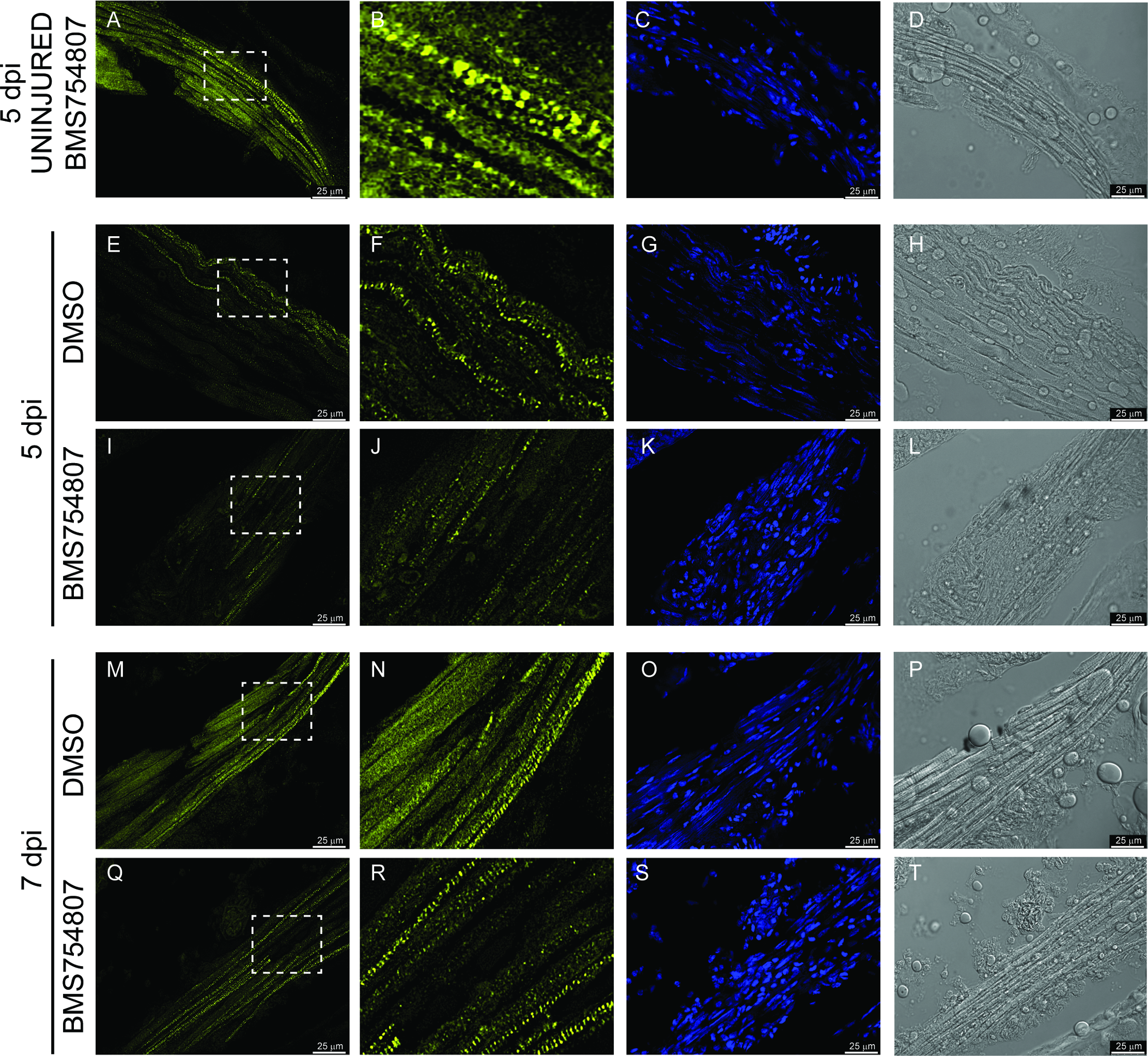
Fig. 5 Myosin staining of the regenerating muscle.
The effect of Igf signaling inhibition on EOM regeneration was analyzed using myosin expression as a marker of muscle differentiation. Uninjured EOMs of BMS754807 treated fish showed high levels of myosin staining (A, B), that were not different of those of a DMSO treated control fish. Myosin staining (yellow) of DMSO control fish at 5 dpi (E, F) and 7 dpi (M, N) reveal higher protein levels than the myosin staining of BMS754807 treated fish at 5 dpi (I, J) and 7 dpi (Q, R). The dashed box shows the approximate position of the magnification shown in B, F, J, N and R. DAPI staining of the corresponding myosin staining picture (C, G, K, O and S) and DIC images (D, H, L, P and T) are also shown. DAPI staining shows hypercellularity in BMS754807 (K, S compared to C, G, O) and typical elongated muscle nuclei in DMSO 7 dpi (O) and Uninjured EOMs (C). Pictures are representative examples of 7 fish per group, time and treatment.