
Image
|
Figure Caption
Fig. S4
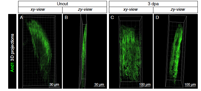
Actinotrichia fibers form palisades on both sides of the distal blastemal mesenchyme.
(A-D) 3D projections of actinotrichia (And1; green) in uninjured (A, B) and at 3 dpa (C, D) fins. Images extracted from S1 Movie. Side views (B, D) show that actinotrichia fibers form two-sided palisades.
Acknowledgments
This image is the copyrighted work of the attributed author or publisher, and
ZFIN has permission only to display this image to its users.
Additional permissions should be obtained from the applicable author or publisher of the image.
Reprinted from Developmental Biology, 433(2), König, D., Page, L., Chassot, B., Jaźwińska, A., Dynamics of actinotrichia regeneration in the adult zebrafish fin, 416-432, Copyright (2017) with permission from Elsevier. Full text @ Dev. Biol.