
Fig. 2
|
Fig. 2
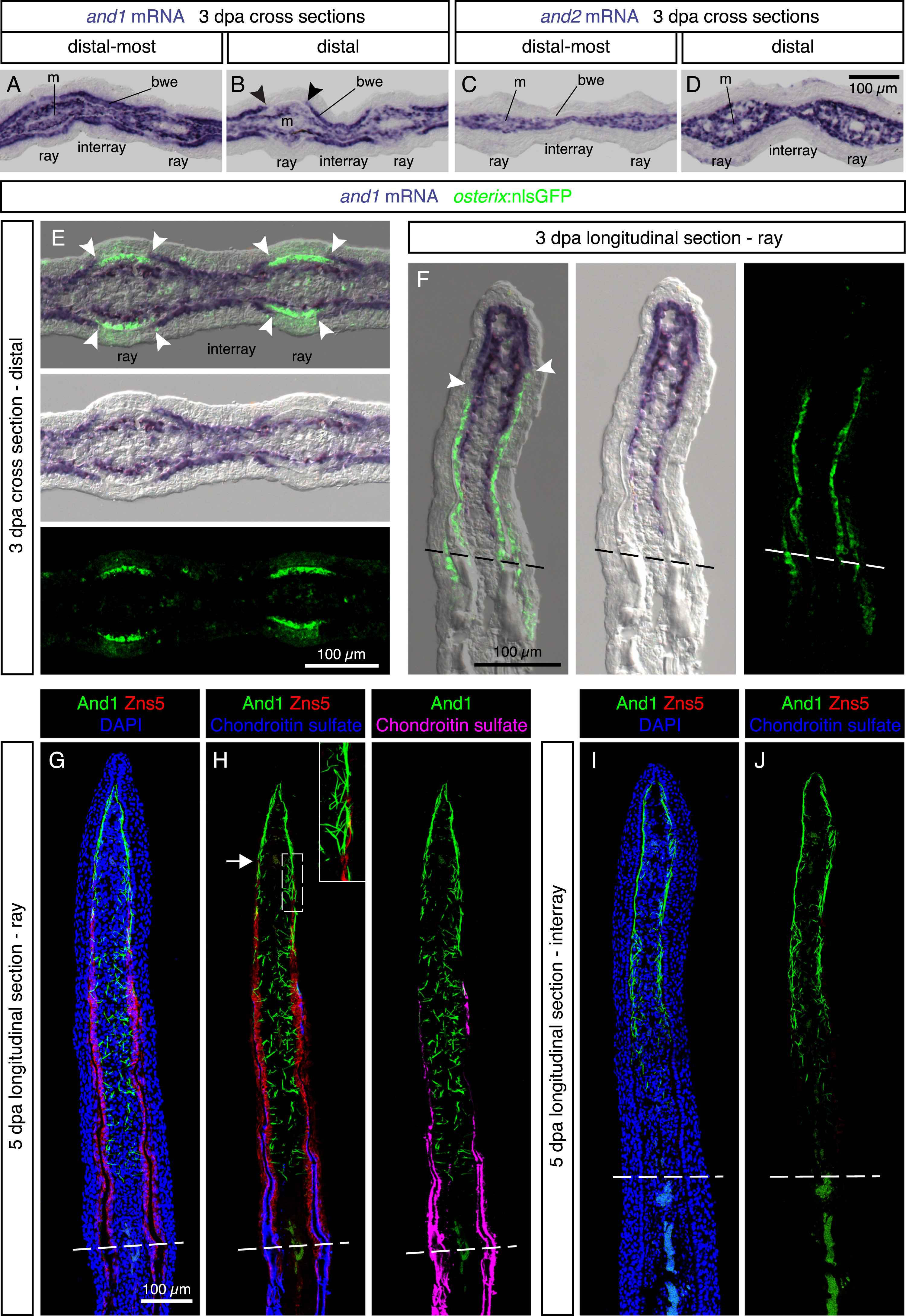
Regenerating lepidotrichia replace actinotrichia in the subepidermal space. (A-D) In-situ hybridization on cross sections of distal-most and distal fin outgrowth at 3 dpa. N = 4 fins. (A, B) In the distal blastema, and1 is differentially expressed in the basal wound epithelium of the rays and interrays. Arrowheads indicate the positions of the interrupted and1 expression in the basal epithelium. m, mesenchyme; bwe, basal wound epithelium. (C, D) and2 is absent from the wound epidermis. (E, F) In-situ hybridization against and1 on transversal (E) and longitudinal sections (F) of osterix:nlsGFP transgenic fins. A lack of and1 expression in the basal wound epithelium of rays is associated with the presence of underlying osteoblasts (green). Arrowheads indicate the positions of the interrupted and1 expression in the basal epithelium. N = 6 fins, 2 sections per fin. (G-J) Immunofluorescence staining for And1 (green), Zns5 (red) and Chondroitin sulfate (H, J, blue) at 5 dpa. (G, H) In the ray, the leading edge of osteoblasts (arrow) replaces actinotrichia in the subepidermal compartment. In the mesenchyme, actinotrichia are disrupted and misaligned. (I, J) In the interrays, no Zns5 and chondroitin sulfate is detected. Actinotrichia remain longitudinally organized in the junctional region between the epidermis and the mesenchyme. The green signal at the base of the outgrowth corresponds to blood autofluorescence. N = 4 fins; 2 sections per fin.
Reprinted from Developmental Biology, 433(2), König, D., Page, L., Chassot, B., Jaźwińska, A., Dynamics of actinotrichia regeneration in the adult zebrafish fin, 416-432, Copyright (2017) with permission from Elsevier. Full text @ Dev. Biol.