
Image
|
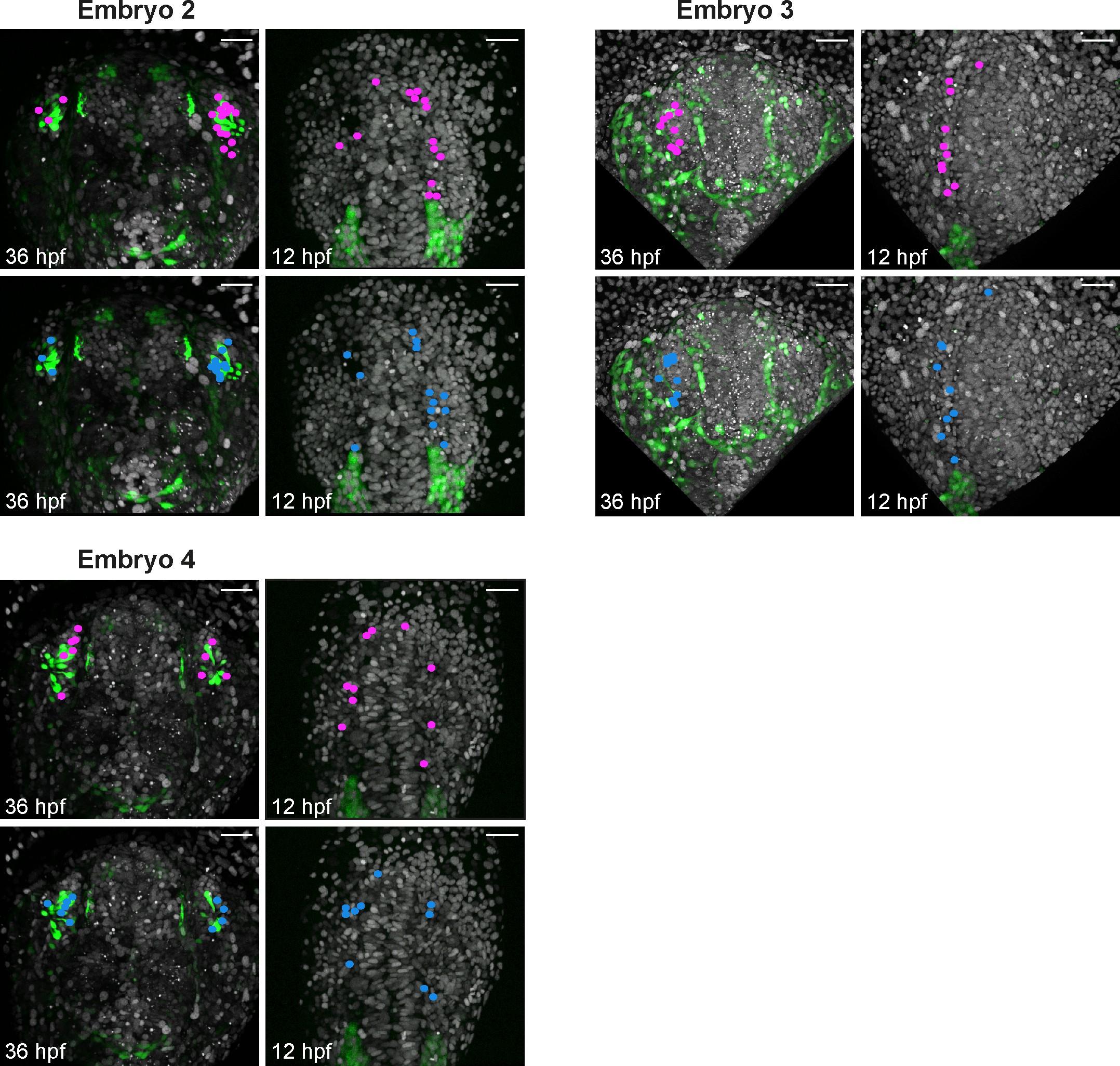
Figure Caption
Fig. 5-S2
Backtracking data from individual Tg(sox10:eGFP) embryos.
Confocal projections extracted from 4D datasets at 36 and 12 hpf for 3 embryos analysed and not shown in Figure 5 showing the position of the backtracked nuclei of sox10:eGFP-negative (pink) and sox10:eGFP-positive (blue) cells at both timepoints. Backtracked nuclei for the lens were only performed for Embryo 1 and are not shown. Embryos are shown with anterior up. Scalebars represent 40 μm.
Acknowledgments
This image is the copyrighted work of the attributed author or publisher, and
ZFIN has permission only to display this image to its users.
Additional permissions should be obtained from the applicable author or publisher of the image.
Full text @ Elife