
Image
|
Figure Caption
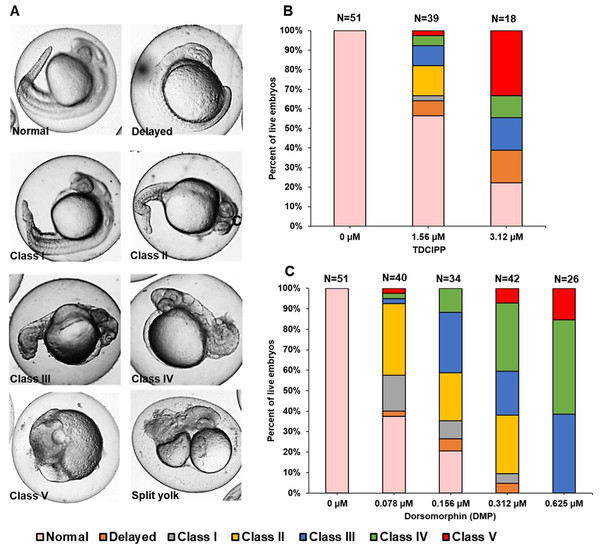
Fig. 2
TDCIPP-exposed embryos phenocopy embryos exposed to dorsomorphin from 0.75 to 24 hpf.
(A) Representative 24-hpf images of normal embryos, delayed embryos, Class I–V levels of dorsalization, and split yolk embryos following exposure to TDCIPP or DMP. (B) Distribution of dorsalization classes following exposure to TDCIPP from 0.75 to 24 hpf. (C) Distribution of dorsalization classes following exposure to DMP from 0.75 to 24 hpf. Depending on the TDCIPP concentration (1.56 or 3.12 µM), the severity of dorsalization varied from mild (Class I) to strong (Class V).
Acknowledgments
This image is the copyrighted work of the attributed author or publisher, and
ZFIN has permission only to display this image to its users.
Additional permissions should be obtained from the applicable author or publisher of the image.
Full text @ Peer J.