
|
Fig. 4
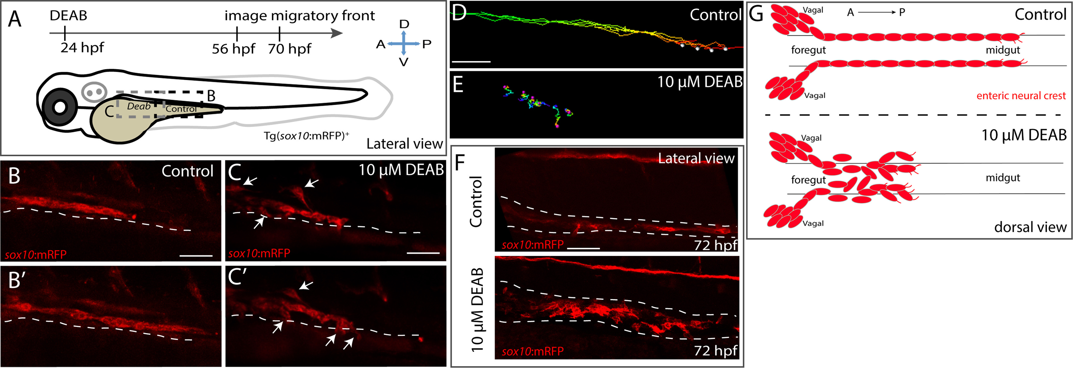
Temporal inhibition of RA results in stalled migration of enteric neural crest chains and loss of chain formation along the foregut. (A) Cartoon schematic of a zebrafish larval fish to illustrate the timing and location of live imaging experiments in panels B-C. Enteric neural crest migratory front cells along the leading edge were imaged in control and experimental conditions. (B-B’,C-C’) Cropped time lapse stills over the course of 3 h showing the enteric neural crest chain front along the foregut-midgut of a sox10:GFP+ control larval fish (B-B’) and the foregut a DEAB treated (C-C’) larval fish. The DEAB treated enteric neural crest chain exhibits delayed migration along the foregut and solitary cells detached from the chain (arrows), while the control enteric neural crest chain front was observed migrating along the midgut collectively. (D-E) Cell tracks of control (D) and DEAB treated (E) enteric neural crest show that DEAB treated enteric neural crest cells fail to progress caudally along the gut, when compared with control. (F) Whole mount immunochemistry against RFP reveals that at 72 hpf, control enteric neural crest chains are maintained along the gut, while DEAB treated enteric neural crest have dissociated along the foregut. (G) Cartoon schematic summarizing the neural crest chain migration phenotype observed following loss of RA along the gut. Red cells depict neural crest migrating in chains along the gut tube in control (top) and in DEAB treated fish (bottom). DEAB treated neural crest chains never make it past the foregut, where they become ectopically localized in the vicinity surrounding the gut. Scale bar in B,C,D and E: 40 μM; scale bar in F: 50 μM.
Reprinted from Developmental Biology, 433(1), Uribe, R.A., Hong, S.S., Bronner, M.E., Retinoic acid temporally orchestrates colonization of the gut by vagal neural crest cells, 17-32, Copyright (2017) with permission from Elsevier. Full text @ Dev. Biol.