
|
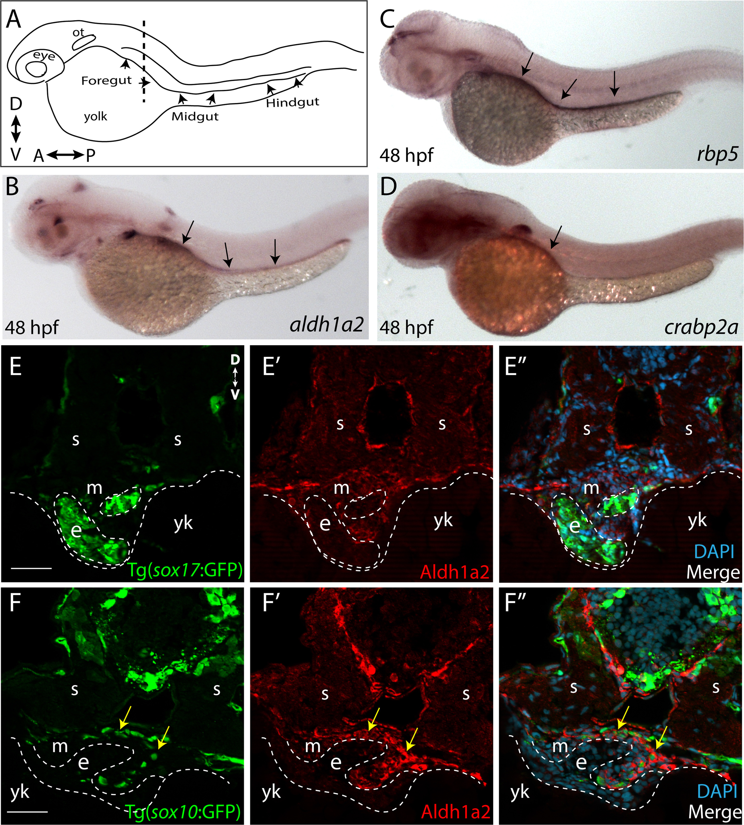
Fig. 1
RA pathway components are expressed along the gut during ENS development. (A) Cartoon illustration of a 1-dpf zebrafish embryo depicted laterally to reveal the location of the foregut, midgut and hindgut. ot-otic, d-dorsal, v-ventral, a-anterior, p-posterior. (B) Whole-mount in situ hybridization against aldh1a2 at 48 hpf reveals its localization along all levels of the gut tube. (C) Whole-mount in situ hybridization against rbp5 at 48 hpf reveals its localization along all levels of the gut tube. (D) Whole-mount in situ hybridization against crabp2a at 48 hpf reveals its localization along the foregut. (E-E’’) Cryosection through the level of the foregut depicts Aldh1a2 protein localization (red) in the gut mesenchyme (m), but not in the gut endoderm (e), marked by sox17:GFP. s-somite, yk-yolk (F-F’’) Cryosection through the level of the foregut depicts Aldh1a2 protein localization (red) in the gut mesenchyme (m) and within neural crest cells migrating in the gut mesenchyme (arrows), marked by sox10:GFP. s-somite, yk-yolk. Scale bars: 50 μM.
Reprinted from Developmental Biology, 433(1), Uribe, R.A., Hong, S.S., Bronner, M.E., Retinoic acid temporally orchestrates colonization of the gut by vagal neural crest cells, 17-32, Copyright (2017) with permission from Elsevier. Full text @ Dev. Biol.