
Fig. 3
- ID
- ZDB-IMAGE-180418-8
- Antibodies
- Publication
- Goody et al., 2017 - Influenza A Virus Infection Damages Zebrafish Skeletal Muscle and Exacerbates Disease in Zebrafish Modeling Duchenne Muscular Dystrophy
- All Figures
- Figures for Goody et al., 2017
|
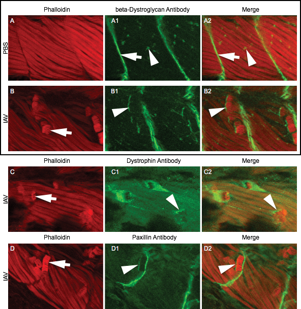
Fig. 3 IAV infection disrupts muscle fiber-ECM adhesion All embryo images are side mounts, dorsal top, anterior left of zebrafish at 24 hpi. Lettered panels show phalloidin staining for F-actin in red. Panels numbered 1 show immunohistochemistry for beta-Dystroglycan, Dystrophin, or Paxillin proteins in green. Panels numbered 2 are merged images of phalloidin and antibody staining. (A-A2) Phalloidin and beta-Dystroglycan staining in a PBS-injected zebrafish. White arrow in A1 denotes MTJ localized beta-Dystroglycan and white arrowhead in A1 points to neuromuscular junction localized beta-Dystroglycan. (B-B2) Phalloidin and beta-Dystroglycan staining in an IAV-injected zebrafish. White arrow in B points to a retracted fiber and white arrowheads in B1-2 highlight beta-Dystroglycan staining at the unattached end of a retracted fiber. (C-C2) Phalloidin and Dystrophin staining in an IAV-injected zebrafish. White arrow in C points to a retracted fiber and white arrowheads in C1-2 highlight Dystrophin staining at the unattached end of a retracted fiber. (D-D2) Phalloidin and Paxillin staining in an IAV-injected zebrafish. White arrow in D points to a retracted fiber and white arrowheads in D1-2 highlight Paxillin staining at the unattached end of a retracted fiber. These results showing that some retracted fibers retain the localization of ECM adhesion proteins suggest that muscle fibers-ECM adhesion can be disrupted external to the sarcolemma.