
Image
|
Figure Caption
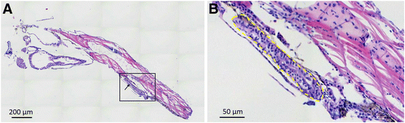
Fig. 4
Histological hematoxylin and eosin (H & E) staining of the whole-mount zPDX model at 7 dpi. A low (a) and a higher (b) magnification of a representative zPDX were showed. Black box in (A) indicates the area of zoom. Arrows in (A) and yellow dashed line in (B) point to the adenoid structure formed by primary epithelial cells from GC patient. Dpi: days post injection
Acknowledgments
This image is the copyrighted work of the attributed author or publisher, and
ZFIN has permission only to display this image to its users.
Additional permissions should be obtained from the applicable author or publisher of the image.
Full text @ J. Exp. Clin. Cancer Res.