
|
Fig. 5
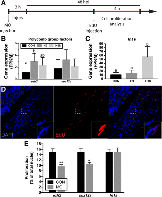
Proliferation following knockdown of select differentially expressed genes. a LR regeneration involves a proliferative burst that generates enough cells to replace lost tissue. EdU assays were performed according to the schematic. b Gene expression (FPKM) of epigenetics selected genes, ezh2 and suz12a. c Fibronectin 1a gene expression (FPKM). d Confocal microscopy of cell proliferation in the regenerating muscle. Inset shows higher resolution detail of the box in the panel. DAPI staining (blue) shows the total number of nuclei in the muscle (left) and EdU staining (red) shows proliferating nuclei (middle). Merged panel (right). e Quantification of cell proliferation in injured LR at 48 hpi. Values are averages ± SEM (n ≥ 5) in control MO or target gene MO injected fish. Different letters (a, b, ab) in (b) and (c) indicate significant differences among time points. * P < 0.05; ** P < 0.01; Student’s t-test