
Image
|
Figure Caption
Fig. s1
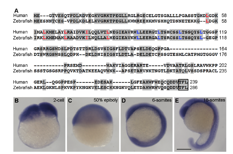
(A) Alignment of human and zebrafish Lurap1 protein sequences. Conserved residues are shadowed. The leucine (L) and isoleucine (I) residues in the tandem leucine repeats are indicated in red and blue, respectively. The C-terminus PDZ-binding motif is boxed. The numbers on the right indicate amino acid positions. (B-E) In situ hybridisation analysis of lurap1 expression pattern in zebrafish embryos at indicated stages. Scale bar: (B-E) 200 μm.
Figure Data
Acknowledgments
This image is the copyrighted work of the attributed author or publisher, and
ZFIN has permission only to display this image to its users.
Additional permissions should be obtained from the applicable author or publisher of the image.
Full text @ Nat. Commun.