
Image
|
Figure Caption
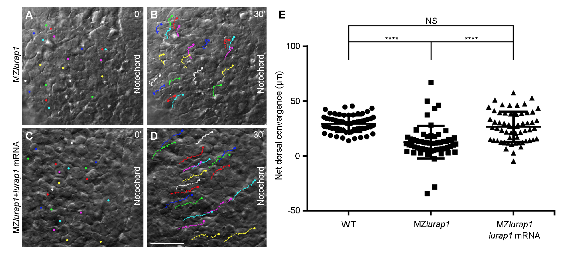
Fig. s5 Lurap1 rescues dorsal convergence of lateral cells in MZlurap1 mutants. (A-D) Representative first and last images from live time-lapse movies show the convergence movement of lateral cells in uninjected MZlurap1, and lurap1-injected MZlurap1 embryos, with the anterior region positioned on the top. (E) Scatter plot shows the net distance reached by lateral cells toward the notochord in indicated embryos. Bars represent the mean values ± s.d. from three independent embryos (****, P<0.0001; NS, not significant; Student’s ttest). Scale bar: (A-D) 50 μm.
Acknowledgments
This image is the copyrighted work of the attributed author or publisher, and
ZFIN has permission only to display this image to its users.
Additional permissions should be obtained from the applicable author or publisher of the image.
Full text @ Nat. Commun.