
|
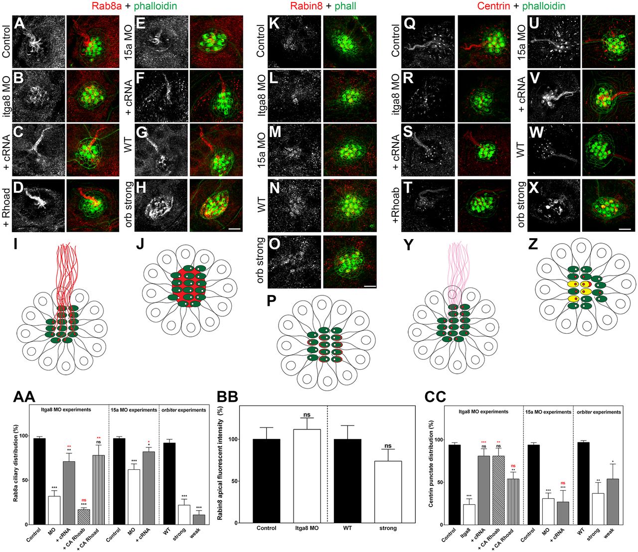
Fig. 7
Lack of the Itga8–Pcdh15a complex activity results in ciliary cargo transport impairment. Confocal images and quantitative data (mean±s.e.m.) for Rab8a (A–H), Rabin8 (K–O) and centrin (Q–X) in control, Itga8 MOs, Pcdh15a MOs and orbiter mutants, and in rescued MOs (labeled as in Fig. 1). Scale bars: 6 µm. (I,J,P,Y,Z) Cartoons of neuromasts (top views) showing the corresponding staining: the hair cell bundle is in green, and Rab8a, Rabin8 or centrin is in red. (I) Control neuromast showing Rab8a ciliary staining. (J) Itga8- or Pcdh15a-deficient neuromast showing apical localization of Rab8a but no staining in the kinocilia. (P) Neuromasts showing apical distribution of Rabin8. (Y) Control neuromast showing basal body/transition zone localization of centrin with weak ciliary staining (pink). (Z) Itga8- or Pcdh15a-deficient neuromast showing centrin localization at the basal body/transition zone and also at the hair cell bundle (yellow). (AA) Rab8a graph. The presence of ciliary Rab8a was evaluated for each treatment in five independent experiments and expressed as percentage of that in control. (BB) Rabin8 graph. Neuromast apical fluorescence was quantified for each treatment in three independent experiments and is expressed as percentage of that in control. (CC) Centrin graph. The centrin punctate distribution correlating with the point of insertion of the kinocilium (basal body/centriole) was qualitatively assessed and the results expressed as percentages of that in control. Five independent experiments were performed. *P<0.05; **P<0.01; ***P<0.001; ns, not significant (one-way ANOVA followed by Dunnett's multiple comparisons test or two-tailed Student's t-test).