
|
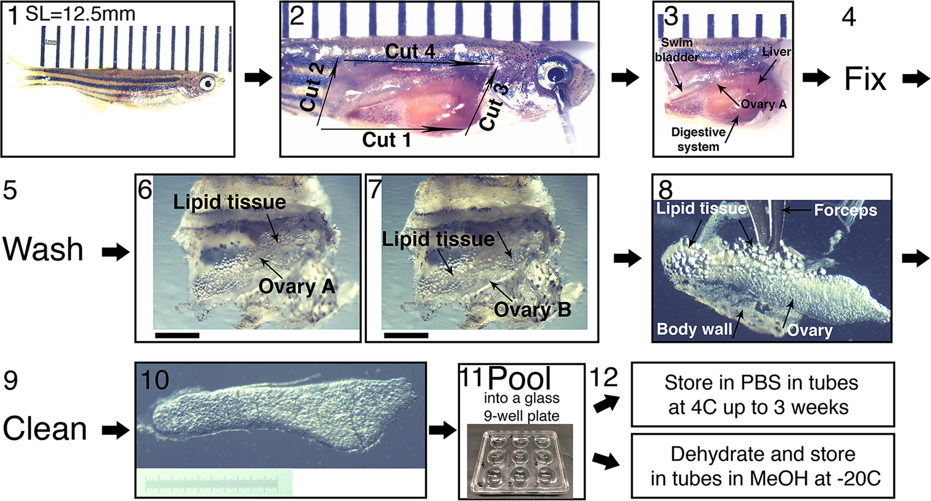
Fig. 2
Collection of ovaries from juvenile fish and their fine dissection. (1) SL measurement. A representative 5 wpf fish of SL=12.5 mm is shown. Fish is laid on a ruler; bars are 1 mm apart. The image is stitched from two smaller frames. (2) The four initial dissection cuts from step 4 in the protocol, exposing the fish internal organs. Bars as in (1). (3) The head and tail are removed and the entire trunk piece is fixed. If ovaries are removed from the fish pre-fixation, they curl up. Fixing the entire trunk piece preserves the ovary morphology. The ovary closer to the image plane (ovary A) and other organs are indicated with arrows. Bars as in (1). (4) The trunk piece is fixed overnight and (5), washed 2-3x in 1xPBS, and the ovaries are finely dissected (6−10). (6−7) The swim bladder, liver and digestive system, are removed from the trunk piece, exposing the ovaries and surrounding lipid tissue. (6) is focused on ovary A and (7) on ovary B. Scale bars are 1 mm. (8−9) The ovaries are removed from the fish, and are cleaned from all lipid tissue and body wall remnants (arrows). (10) An example of a clean ovary. Scale bar is 1 mm (graded 10 µm). (11) Ovaries can be pooled in a glass well-plate and (12) stored in the appropriate conditions for the subsequent application.
Reprinted from Developmental Biology, 430(2), Elkouby, Y.M., Mullins, M.C., Methods for the analysis of early oogenesis in zebrafish, 310-324, Copyright (2017) with permission from Elsevier. Full text @ Dev. Biol.