
|
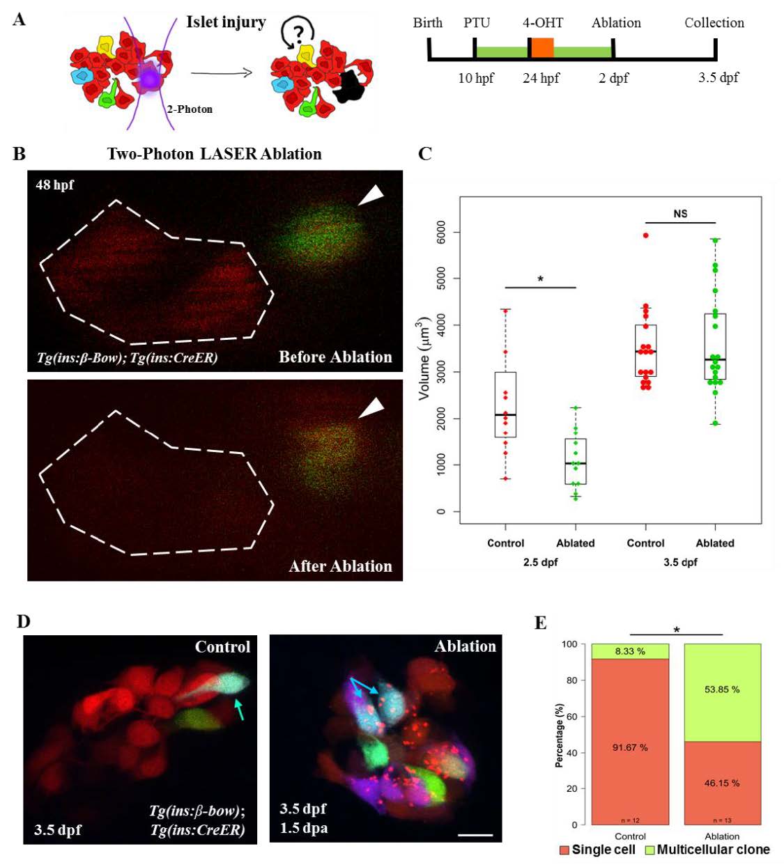
Fig. S8
Injury based reversal of DBCs quiescence. (A) Cartoon (left) and a schematic (right) for the two-photon laser-injury model. The model tests if partial islet-ablation is capable of inducing a proliferative response among the quiescent dorsal bud-derived beta-cells (DBCs). (B) Snapshots from in-vivo imaging of 48 hpf beta-bow animals undergoing two-photon laser-ablation. DBCs were labeled in multiple colors at 24 hpf by 4-OHT-incubation. The dotted line indicates the target area, composed of un-recombined cells (red). Arrowheads point to recombined cells, located outside of the target area. These cells were not ablated. (C) Quantification of the total volume of the beta-cells in the primary islet following laser-ablation. The plot shows tukey style boxplot overlaid with the data points. At 12 hours post-ablation (2.5 dpf), the ablated islets exhibited smaller volumes compared to the control islets (unpaired t-test,* p ≤ 0.05). At 36 hours post-ablation (3.5 dpf), the volume of beta-cells in the ablated samples was similar to controls. (D) Maximum intensity projections of control and ablated primary islets. Arrows indicate trichromatic cells. Note the presence of red puncta in the beta-cells from the laser-ablated islets, which might indicate aggregation of misfolded proteins due to induction of cellular stress after the injury. (E) Quantification of the proportion of trichromatic beta-cells that remain as single cells or form multicellular clones in the control and laser-ablated islets. Multicellular clones denote at least two cells per clone. Approximately one-half of DBCs underwent cell divisions in response to injury, a statistically significant increase in proliferation compared to controls (Fisher’s exact test,* p ≤ 0.05). Scale bars, 10 μm