
Image
|
Figure Caption
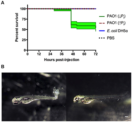
Fig. 2
Zebrafish larvae exposed to P. aeruginosa by injection. Larvae were injected at 72 HPF with 2,000–6,000 CFU of P. aeruginosa PAO1 or E. coli DH5α into the caudal artery. Sterile PBS was injected as control. (A) Survival curve of 3 DPF larvae injected with P. aeruginosa PAO1 grown in PGS (↓Pi) medium (green line), P. aeruginosa PAO1 grown in PGS (↑Pi) medium (red dashed line), E coli DH5α (blue line) or sterile PBS medium (black dotted line). (B) Larvae injected at 72 HPF with P. aeruginosa PAO1 grown in PGS (↓Pi) medium (left) or injected with sterile PBS medium (right) at 28 HPI. Scale: 100 μm.
Figure Data
Acknowledgments
This image is the copyrighted work of the attributed author or publisher, and
ZFIN has permission only to display this image to its users.
Additional permissions should be obtained from the applicable author or publisher of the image.
Full text @ Front Cell Infect Microbiol