
|
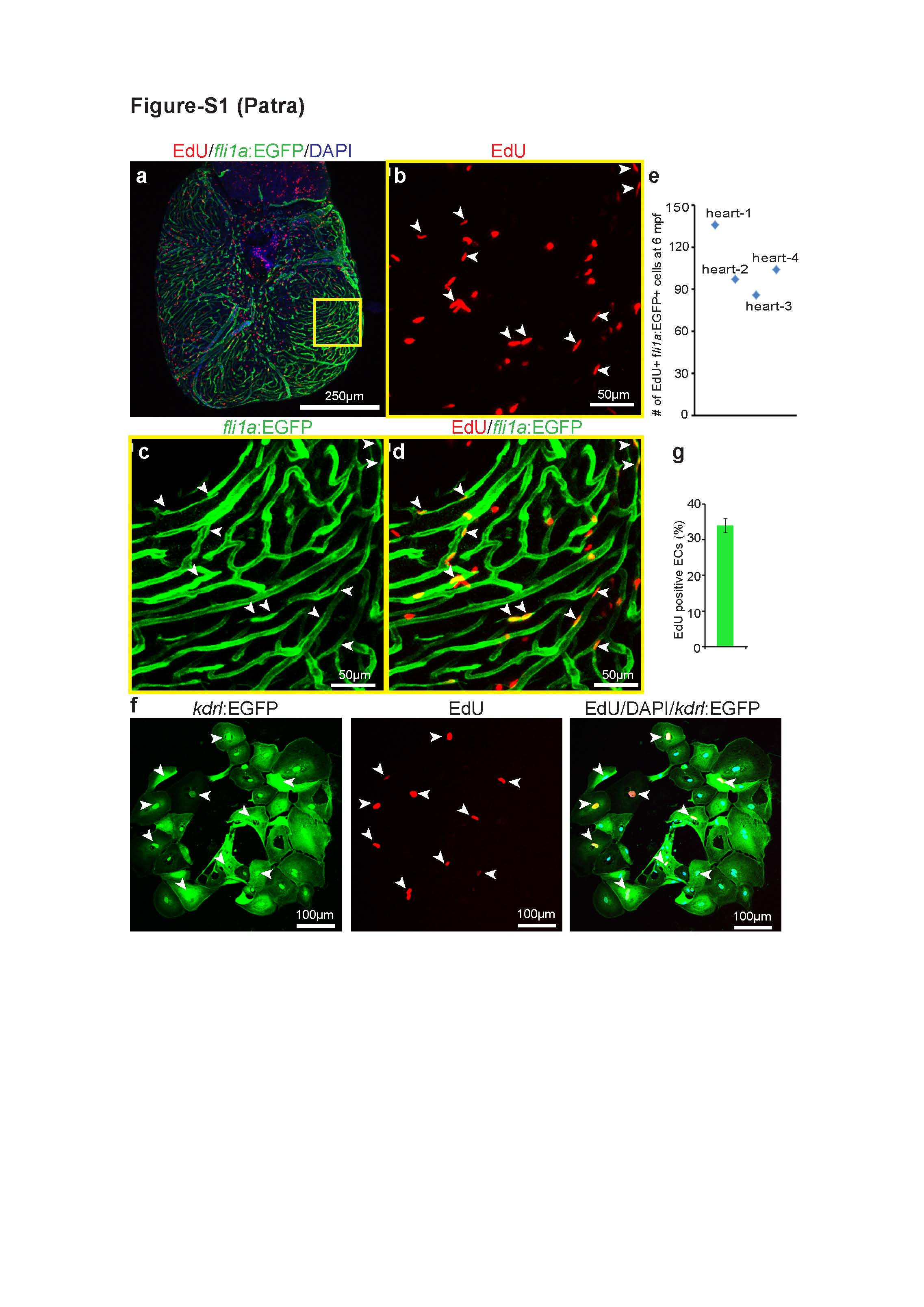
Fig. S1
Cardiac endothelial cells are proliferative in vivo. (a) Representative views of maximum confocal projections through 6 months old Tg(fli1a:EGFP) zebrafish whole mount hearts stained for EGFP (green), EdU (red), and DAPI (nuclei, blue). (b-d) Representative confocal images of ‘a’. Arrowheads point to EdU+ EGFP+ cells. (e) Quantification of EdU/EGFP-positive coronary endothelial cells. (n=4). (f) Representative views of cultured endothelial cells stained for EGFP (green), EdU (red), and DAPI (nuclei, blue). White arrowheads point to EdU-positive endothelial cells. (g) Quantification of EdU+ EGFP+ endothelial cells with 10% FBS in culture medium (n=3, mean±SEM).