
|
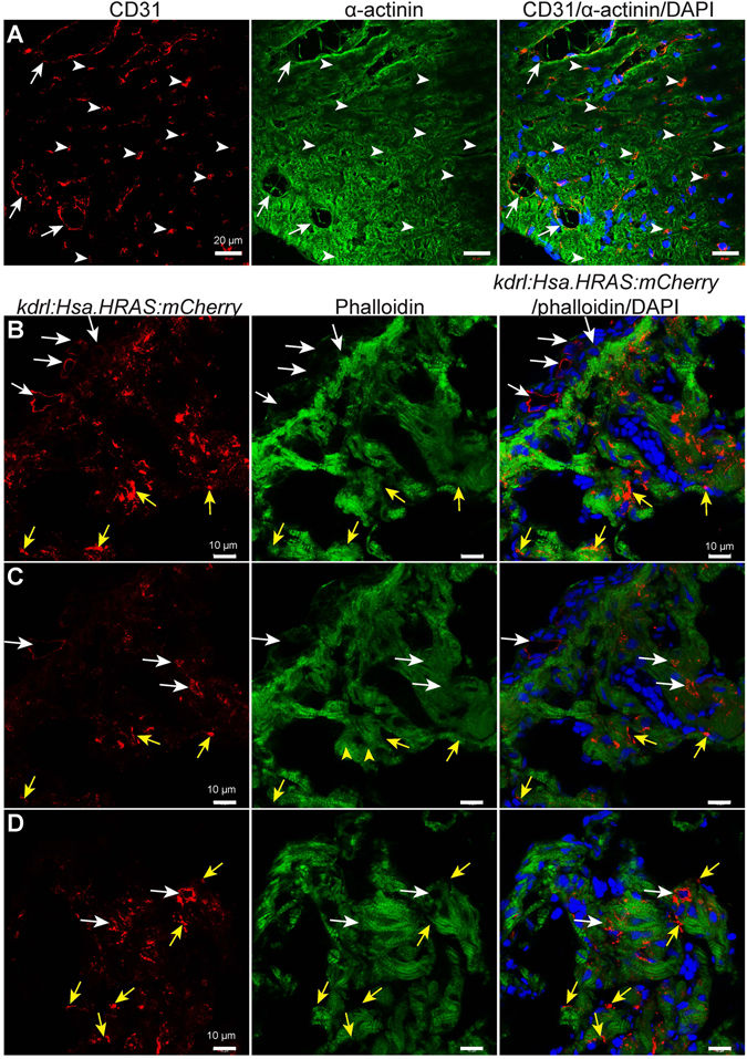
Fig. 3
Endothelial cell distribution in 8 months old mouse and zebrafish ventricles. (A) Representative confocal images of sagittal cryosections through 8 months old mouse hearts stained for α-actinin (green; marking cardiomyocytes), CD31 (red; marking the cell membrane of endothelial cells) and DAPI (blue, staining the nuclei). White arrows and arrowheads point to bigger coronary vessels (visible lumen) and capillaries (lumen not visible) in cardiac tissue, respectively. (B–D) Representative confocal images of sagittal cryosections through 8 mpf Tg(kdrl:Hsa.HRAS-mCherry) zebrafish hearts stained for phalloidin (green; marking cardiac tissue), mCherry (red; marking the cell membrane of endothelial cells) and DAPI (blue; staining the nuclei). White arrows point to bigger coronary vessels (visible lumen). Yellow arrows point to endocardial cells in the cardiac tissue. ‘B’ and ‘C’ images show different optical planes of the same tissue section.