
|
Fig. S7
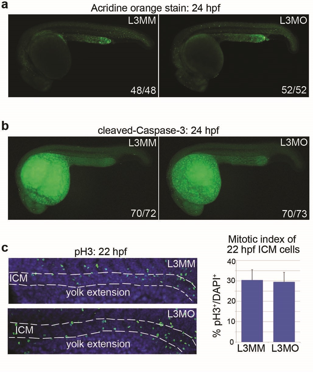
Knockdown of drl.3 does not alter cell survival or proliferation during embryonic hematopoiesis.
(a) Acridine orange staining of L3MMand L3MO-injected embryos at 24 hpf. {b) lmmunofluorescent detection of cleaved Caspase-3 in L3MMand L3MO-injected embryos at 24 hpf. (a-b) Numbers of embryos with representative phenotypes per total embryos analyzed are indicated. (c) Confocal analysis of phospho-Histone H3 (pH3, green) immunodetection and DAPI (blue) staining in L3MM- and L3MO-injected embryos at 22 hpf. Dashed line outlines the ICM. The mitotic index of ICM cells is shown in the right panel. Bars are mean ± S.D. from 8 embryos per condition.