
Image
|
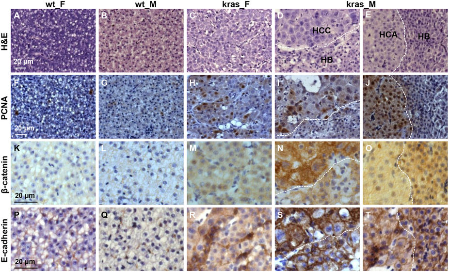
Figure Caption
Fig. 4
Characterization of the multi-nodular liver tumor from male krasV12 zebrafish after long-term tumor induction.
Adjacent liver sections in female and male wild type and krasV12 fish at 5 mpi were processed for H&E and immunohistological staining. The multi-nodular male tumors have mixed types of tumors (HCC, HB and HCA) which were separated by dash lines. (A–E) H&E staining. (F–J) Immunocytochemical staining for PCNA. (K–O) Immunohistochemical staining for β-catenin. (P–T) Immunohistochemical staining for E-cadherin. Scale bar, 20 μm for all panels.
Figure Data
Acknowledgments
This image is the copyrighted work of the attributed author or publisher, and
ZFIN has permission only to display this image to its users.
Additional permissions should be obtained from the applicable author or publisher of the image.
Full text @ Sci. Rep.