
Image
|
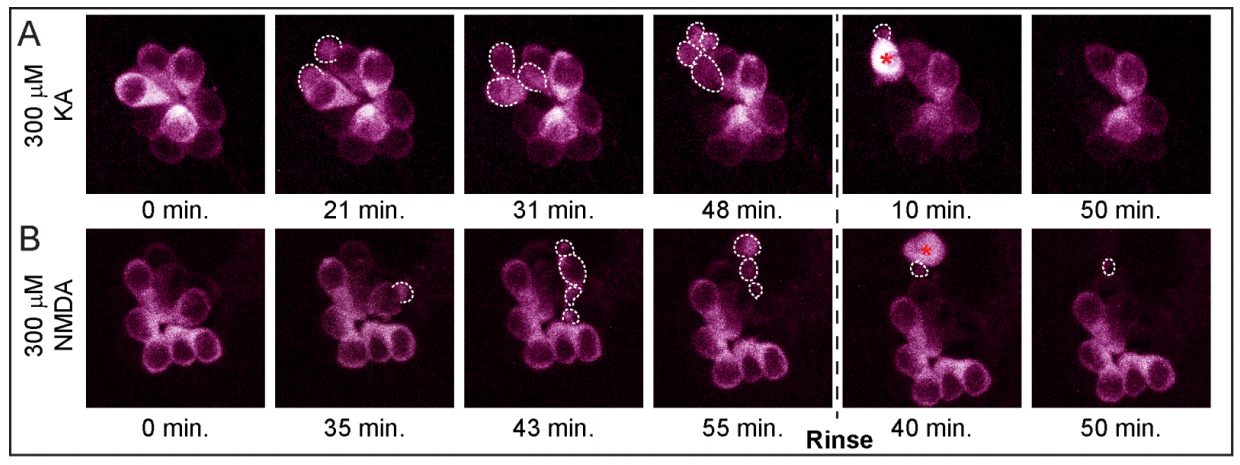
Figure Caption
Fig. S1
A subset of hair cells in KA- and NMDA-exposed NMs form apoptotic bodies prior to cell death.
A-B) Max projection z-stack images from live-imaging of an NM in neurog1 morphant larvae with hair cells stably expressing GCaMP3. Z-stack images were taken every 30 seconds for 2.5 hours total; 300 μM KA (A) or 300 μM NMDA (B) was applied for 50 minutes, then rinsed with E3 media which remained for the duration of imaging. White dashed outlines indicate dying hair-cells blebbing and fragmenting into apoptotic bodies.
Acknowledgments
This image is the copyrighted work of the attributed author or publisher, and
ZFIN has permission only to display this image to its users.
Additional permissions should be obtained from the applicable author or publisher of the image.
Full text @ Sci. Rep.