
|
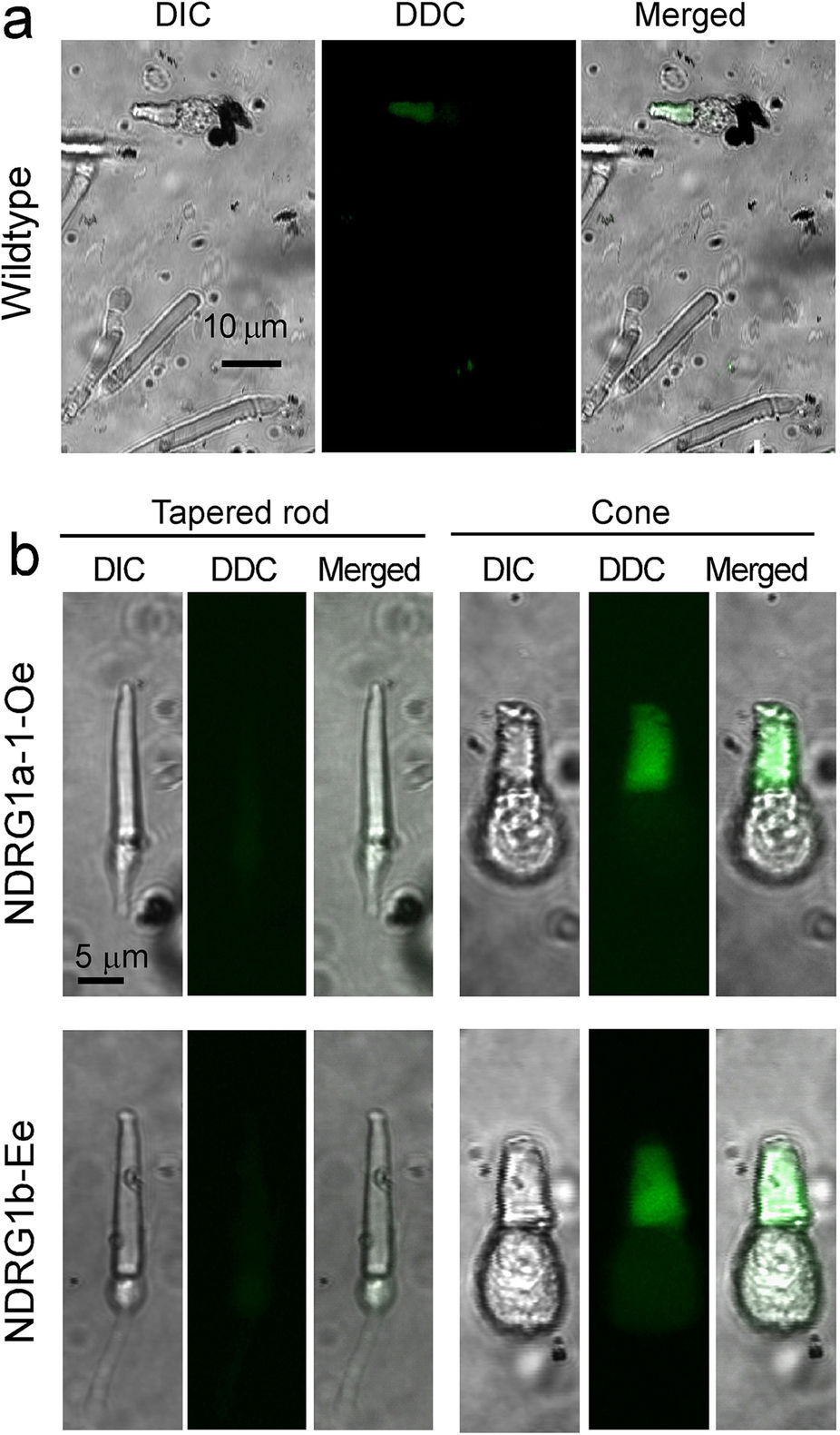
Fig. 5
DDC staining of rods showing taper-shaped OS.
(a) Typical DDC staining of wildtype (Wildtype) rods and cones. Only cone OS was stained. (b) Rods with tapered OS obtained from NDRG1a-1-Oe (NDRG1a-1-Oe) and NDRG1b-Ee (NDRG1b-Ee) was negative for DDC staining (upper and lower left 3 panels, respectively). Cone OSs in NDRG1a-1-Oe and NDRG1b-Ee were positively stained (upper and lower right 3 panels, respectively). Cells in (a,b) were viewed with DIC (DIC) and under fluorescent microscope to detect the fluorescence of DDC (DDC). Scale bar is 10 μm in (a). Magnifications are the same in all of the panels in (b) (scale bar, 5 μm).