
|
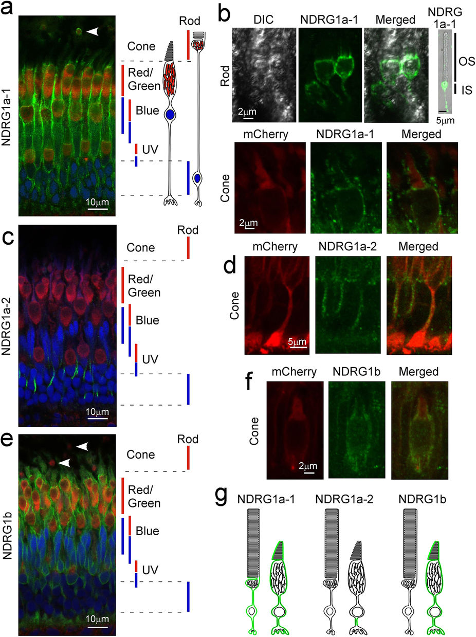
Fig. 1
Subcellular localization of NDRG1 family proteins in zebrafish photoreceptors.
Adult zebrafish retinas were immunostained with specific anti-NDRG1a-1 (a,b), anti-NDRG1a-2 (c,d) or anti-NDRG1b (e,f) antiserum (all with green signals). Mitochondria and nucleus were counterstained with anti-Tom20 antibody (red signals) and Hoechst 33342 (blue signals), respectively (a,c,e). In a, c and e, approximate positions of ellipsoid containing mitochondria (red bars) and nucleus (blue bars) for each cone type (red/green-, blue- and UV-sensitive cones) and for rods are indicated. NDRG1a-1 was found to be expressed in the ellipsoid but not in the OS in rods (arrowhead in a, and upper panels in b). NDRG1a-1 was also found both in the OS and ellipsoid in cones (a and lower panels in b). NDRG1a-2 was found in the thin process of a cone (c,d). NDRG1b was found in the entire region of a cone (e,f) but not in rod ellipsoid (arrowheads in e). In a, c and e, wildtype zebrafish retinas were used, and in the detailed study in b, d and f, the retinas consisting of cones expressing mCherry-HrasCAAX were used to readily identify cones. (g) Schematic representation of localization of NDRG1 family proteins in adult zebrafish photoreceptors (shown in green). DIC, differential interference contrast image.