
Image
|
Figure Caption
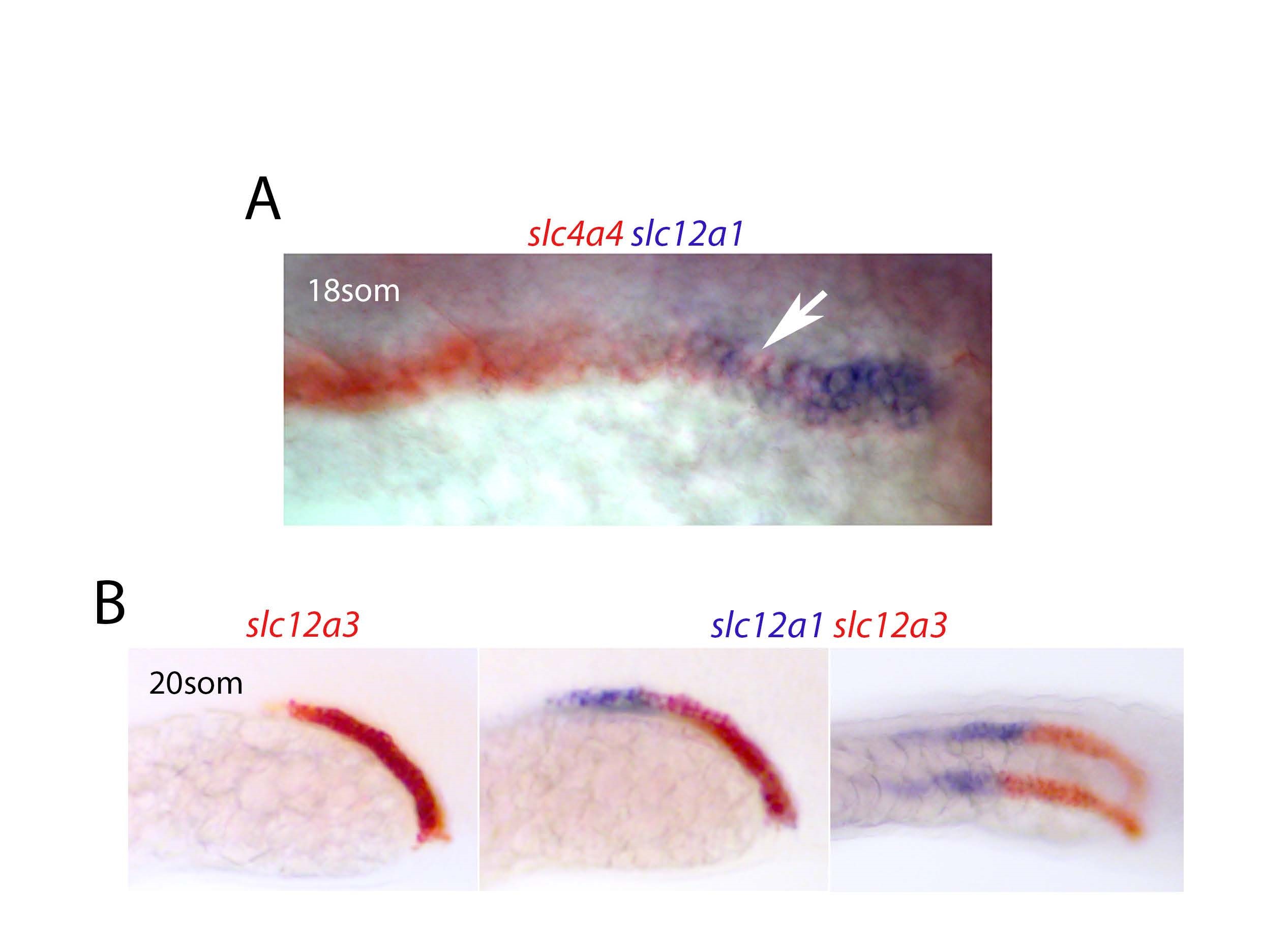
Fig. S1
A) Expression of slc4a4/slc12a 1 at the 18-somite stage. White arrow highlights mixing of these two populations. B) Left panel shows a lateral view of 20-somite stage embryo stained for slc12a3 in red. Middle and right panels show the same embryo but also with slc12a1 staining in purple with the middle panel being a lateral view and the right panel an oblique view.
Acknowledgments
This image is the copyrighted work of the attributed author or publisher, and
ZFIN has permission only to display this image to its users.
Additional permissions should be obtained from the applicable author or publisher of the image.
Full text @ Sci. Rep.