
Image
|
Figure Caption
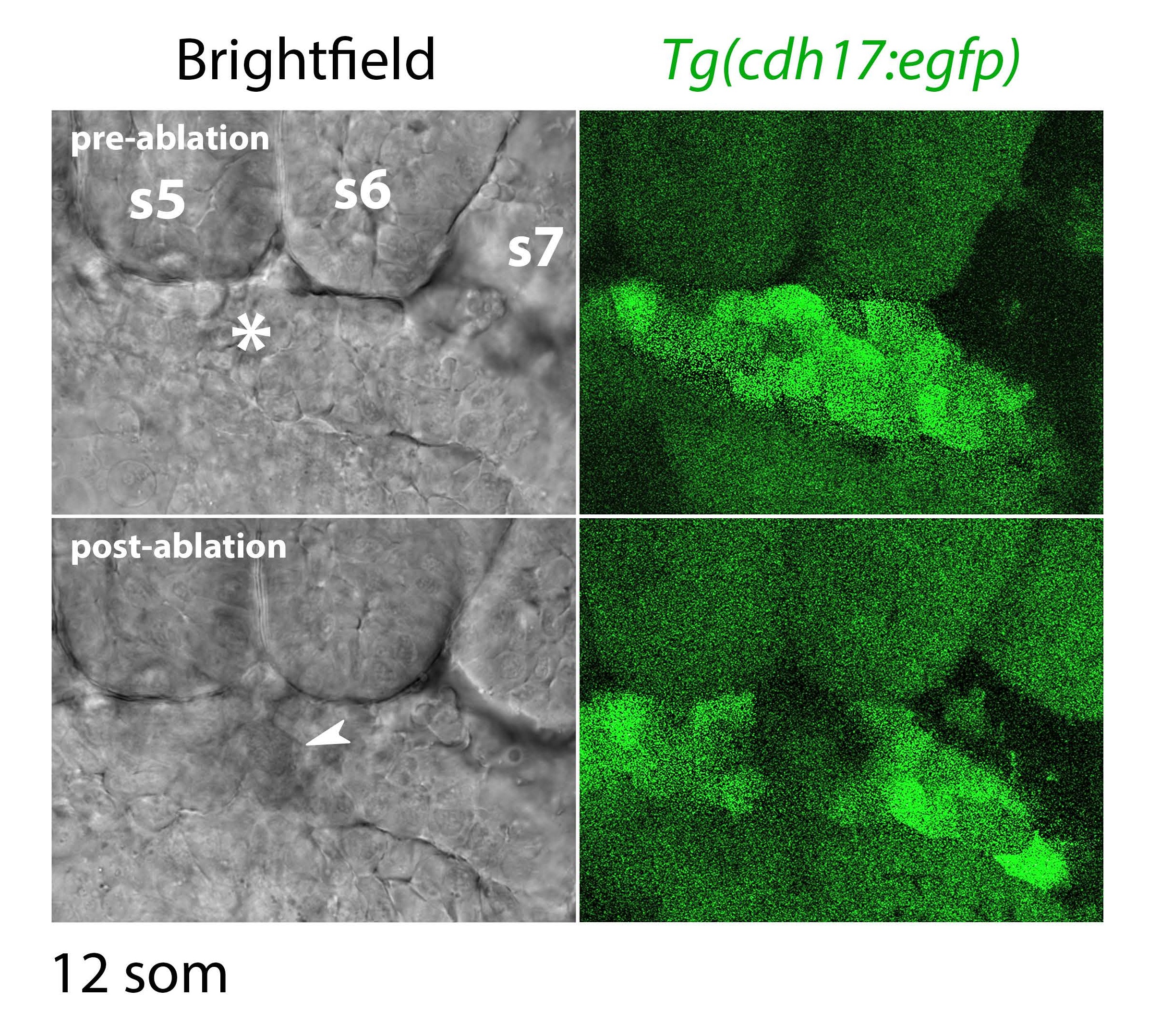
Fig. S7
Laser ablation of the tubule. Panels show close-up images of Tg(cdh 17:egfp) embryos under brightfield and fluorescence microscopy at the 12-somite stage where 405nm wavelength light on a confocal microscope has ablated the intermediate mesoderm (asterisk marks target site for ablation, white arrowhead highlights the target site immediately post-ablation).
Acknowledgments
This image is the copyrighted work of the attributed author or publisher, and
ZFIN has permission only to display this image to its users.
Additional permissions should be obtained from the applicable author or publisher of the image.
Full text @ Sci. Rep.