
|
Fig. 3
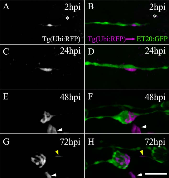
Regenerated neuromasts are chimeric structures derived from two interneuromastic cells (INCs). Transplanting cells from a tg(ubiquitin:RFP) blastula to a tg(et20:GFP) blastula occasionally resulted in fish with one or a few labeled INCs. A transplanted larva harboring a single labeled INC near L3 was selected 3 days post fertilization and subjected to electroablation of the L3 neuromast (a, b). The asterisc indicates the position of the ablated neuromast. The left panels (a, c, e, g; only the red fluorescent protein [RFP] channel is shown) show the behavior of the transplanted cell through time. The right panels (b, d, f, h) show ET20+ cells of host larvae (green) and the transplanted cells (pseudocolored in magenta). During regeneration, the single transplanted cell divided and its progeny differentiated into different cells types of the mature neuromast. At 72 hpi, a red-labeled cell (here in magenta) can be observed among the INCs, suggesting that at least one daughter cell maintained the original identity of the progenitor (g, h, yellow arrowhead). Note that the transplantation experiment randomly generated labeled cells of diverse lineages that did not participate in neuromast regeneration (see for example, e–h, white arrowhead). Scale bar: 50 μm