
|
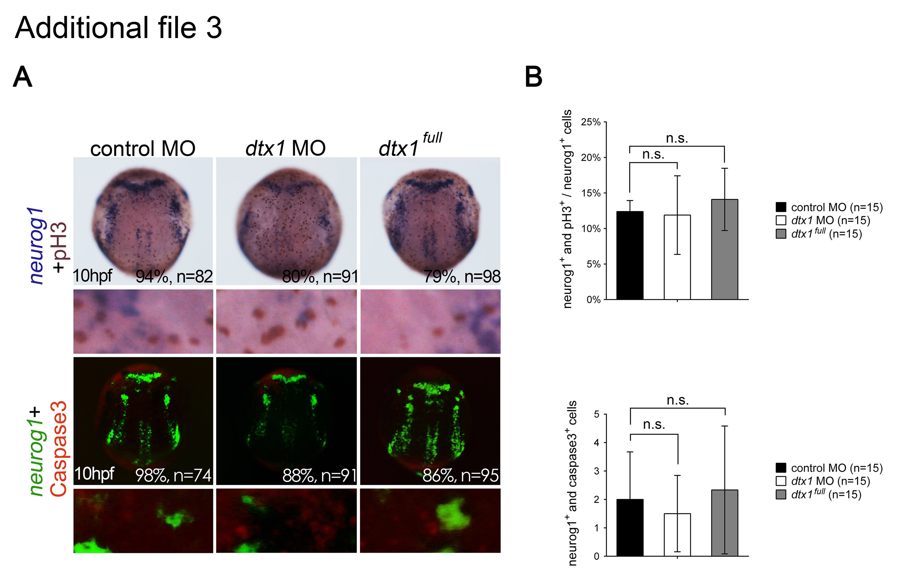
Fig. S3 Disruption of Dtx1 expression had no effects on neuronal proliferation and apoptosis. a. Proliferating neurons were double-labeled with phospho-histone H3 antibody (brown) and neurogenin1 riboprobes (purple). Apoptotic neuronal precursor cells were labeled for neurogenin1 (fluorescent green) and activated caspase-3 antibody (fluorescent red). b. Proliferating neuronal cells were quantified by counting the proportions of phospho-histone H3- and neurogenin1-positive cells, whereas apoptotic neural progenitor cells were quantified by counting the proportions of activated caspase-3- and neurogenin1-positive cells, revealing no marked differences between the embryos injected with dtx1 full , dtx1 morpholinos, and the controls. n.s., nonsignificant.