
|
Fig. 3
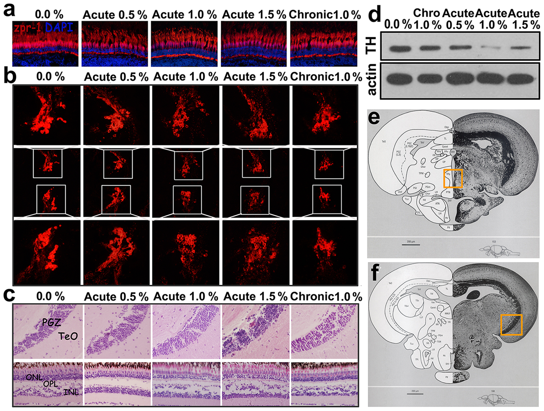
The effects of acute and chronic alcohol exposure on the retina and brain of the zebrafish.
(a) Cone photoreceptor cells labelled by the Zpr-1 antibody in wild-type zebrafish and zebrafish groups treated with alcohol. (b) Tyrosine hydroxylase (TH) expression in the posterior tuberculum (PT) detected by immunohistochemistry and western blotting. (c) The histopathology of the brain and retina in wild-type zebrafish and in the groups treated with alcohol. (d) TH expression in the brain detected by western blotting. (e) The boxed area in the section from the adult zebrafish brain highlighting the brain region shown in B. (f) The boxed area in the section from the adult zebrafish brain highlighting the brain region shown in C. Cropped blots are used in the figure, and full-length blots are presented in Fig J in S1 File. The images in E and F are reproduced from Wullimann, M., Neuroanatomy of the Zebrafish Brain: A topological atlas, Birkhauser press, Basel (1996)36, Chapter 5, Fig 153 and Chapter 5 Fig 168, respectively, with permission from Springer Science and Business Media.