
|
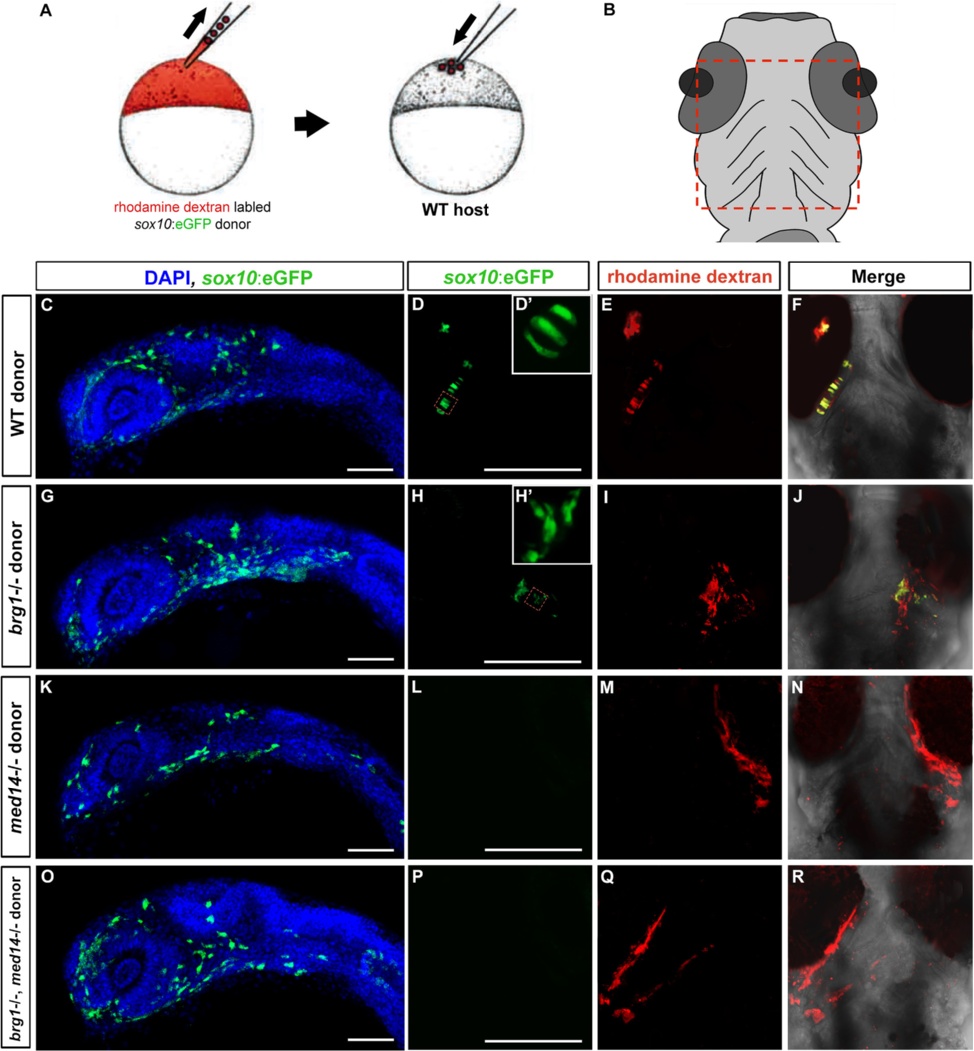
Fig. 7
Cell autonomous requirement for med14 and brg1 in neural crest cells for cartilage differentiation. a Schematic diagram of transplantation approach. Donor (wild type or mutant) sox10:EGFP transgenic embryos were injected with rhodamine-dextran lineage tracer and transplanted to the animal pole of wild type host embryos at 4 hpf. b Diagram showed the region was imaged at 80 hpf. c, g, k and o At 24 hpf, donor-derived neural crest migration to the oral ectoderm is evident regardless of donor genotype. Later views with anterior to the top. d, e and f; h, i and j; l, m and n; p, q and r Donor cell contribution to cartilage assayed at 80 hpf. Ventral views with anterior to the top. D’ and H’: higher magnificent views of regions indicated by red squares in b and e. Scale bars, 100 um