
|
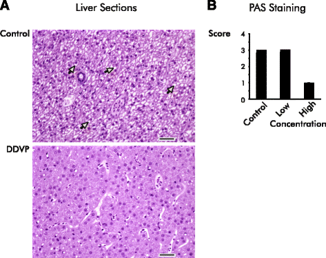
Fig. 2
Glycogen vacuolation is reduced in the livers of DDVP–exposed fish. a The liver of exposed fish shows decreased hepatocellular vacuolation compared with control animals (yellow arrows). Representative images of transversely sectioned hematoxylin and eosin stained livers from a control and a unexposed fish (high concentration) are shown. Bars are 25 μm. b Periodic Acid-Schiff (PAS) staining reveals reduced levels of glycogen in the livers of exposed fish. Liver sections from controls and fish exposed to high and low concentrations of DDVP were semi-quantitatively scored for PAS-positive glycogen staining (0-4+, low to high staining intensity). All unexposed fish were scored “3 + .” Scores for the low concentration-exposed fish ranged from 2 + −4+ (mean = 3+), and all the fish exposed to the high concentration were scored “0–1+”. Exposed fish differed from control at p ≤ 0.05 level using the Kruskal-Wallis test. The high concentration exposure was significantly different from control in post hoc testing with the Wilcoxon ranks test (p ≤ 0.05)