
|
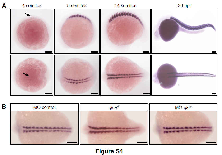
Fig. S4
Related to Figure 4. qkia and qkic are not individually essential for tpm3.12 expression in differentiating slow muscle cells.
(A) tpm3.12 is expressed in differentiating slow muscle cells. tpm3.12 transcripts are first detected by in situ hybridization at 11 hpf (4-somite stage) in slow muscle cells (arrow). Its expression pattern in differentiating slow muscle cells follows the antero-posterior progression of somitogenesis until 26 hpf, when tpm3.12 transcripts are specifically expressed in trunk muscle. Note that embryos at 26 hpf are overstained to ensure that tpm3.12 is not expressed in other cells than muscle cells. Lateral (top panel) and dorsal views (bottom panel) of the same embryo are shown.
(B) tpm3.12 expression is not affected in qkia mutant or qkic morphant embryos. At 14/15 hpf (10/12- somite stage), tpm3.12 expression is similar in sibling and qkia-/- embryos injected with control MO (n=23) as well as in sibling injected with qkic MO (n=40).
In all panels, anterior is to the left. Scale bar 100μm.
Reprinted from Developmental Cell, 42(5), Bonnet, A., Lambert, G., Ernest, S., Dutrieux, F.X., Coulpier, F., Lemoine, S., Lobbardi, R., Rosa, F.M., Quaking RNA-Binding Proteins Control Early Myofibril Formation by Modulating Tropomyosin, 527-541.e4, Copyright (2017) with permission from Elsevier. Full text @ Dev. Cell