
|
Fig. 7
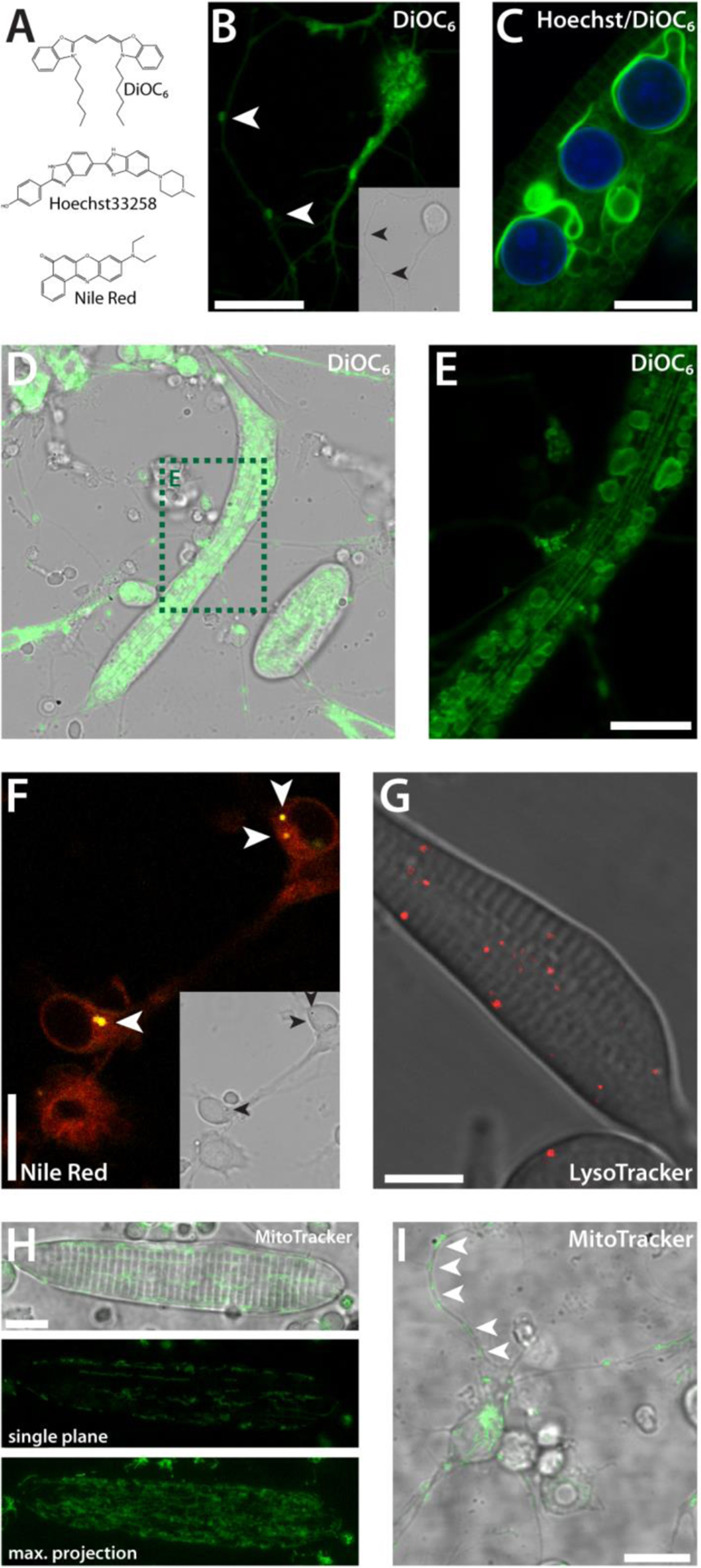
Vital dyes. (A) Chemical structures of commonly used cell-permeant dyes. (B) Neuron-like cell stained with DiOC6 (1 dap). The complete endomembrane system is visible. In a nearby process of a different cell, stained clusters can be observed (white arrowheads). Inset: Corresponding transmitted light image. (C) Striated myocyte co-stained with DiOC6 and Hoechst33258 (1 dap). (D,E) Striated myocyte, stained with DiOC6 (2 dap), (E) is a detail of (D). (F) Neuron-like cells stained with Nile Red (1 dap). Membranes are displayed in red, lipid droplets in yellow (white arrowheads). Inset: Corresponding transmitted light image. (G) Striated myocyte stained with LysoTracker (2 dap). (H) Myocyte stained with MitoTracker (2 dap). (I) Neuron-like cell stained with MitoTracker with distinct mitochondria along processes (2 dap). Scale bars, 10 µm. Each staining method was performed three times or more with several wild type cultures.
Reprinted from Developmental Biology, 430(1), Sassen, W.A., Lehne, F., Russo, G., Wargenau, S., Dübel, S., Köster, R.W., Embryonic zebrafish primary cell culture for transfection and live cellular and subcellular imaging, 18-31, Copyright (2017) with permission from Elsevier. Full text @ Dev. Biol.