
Image
|
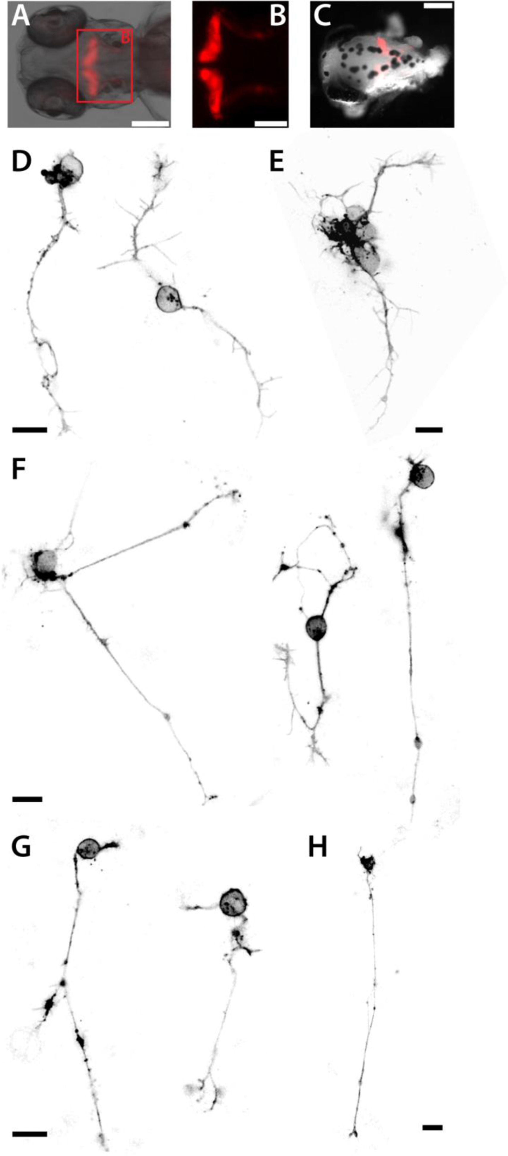
Figure Caption
Fig. 6
Purkinje cells of 4 dpf embryos in culture. (A) Tg(ca8:FyntagRFP)bz4Tg embryo (dorsal view, 4 dpf). The cerebellum with red fluorescent Purkinje cells shown in (B) is marked by a red box. Scale bar, 100 µm. (C) Decapitated head of a 4 dpf Tg(ca8:FyntagRFP)bz4Tg embryo. Scale bar, 100 µm. (D-H) Red fluorescent Purkinje cells displaying characteristic morphologies at 1 (D,E), 2 (F) and 3 (G,H) dap. Scale bars, 10 µm. The figure shows a collection of the images of two preparations with comparable results.
Acknowledgments
This image is the copyrighted work of the attributed author or publisher, and
ZFIN has permission only to display this image to its users.
Additional permissions should be obtained from the applicable author or publisher of the image.
Reprinted from Developmental Biology, 430(1), Sassen, W.A., Lehne, F., Russo, G., Wargenau, S., Dübel, S., Köster, R.W., Embryonic zebrafish primary cell culture for transfection and live cellular and subcellular imaging, 18-31, Copyright (2017) with permission from Elsevier. Full text @ Dev. Biol.