
Image
|
Figure Caption
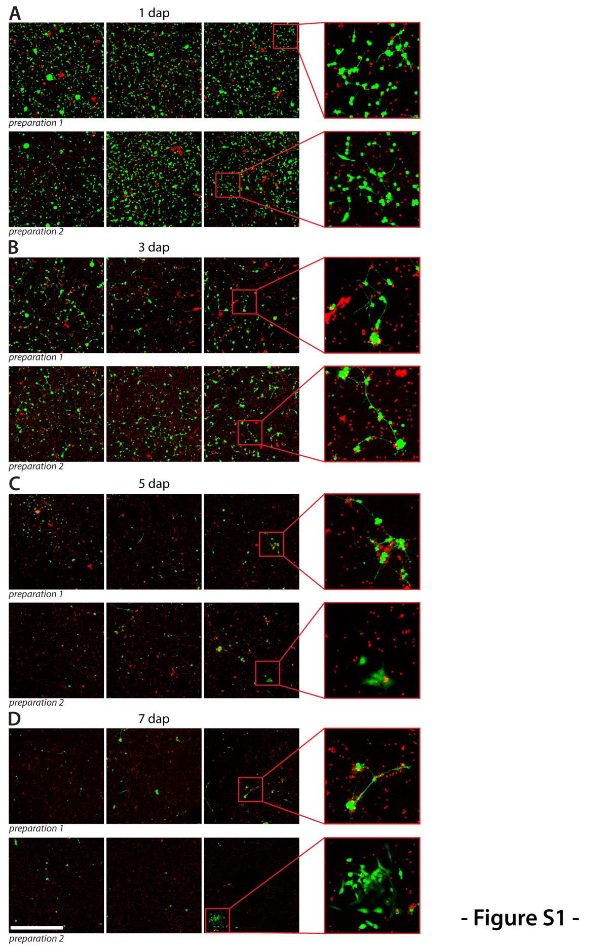
Fig. S1
Survival of primary cells. Two cultures were prepared from wild type embryos. Each day, one dish of each culture was co-stained with Calcein-AM to label live cells (green) and propidium iodide which stains all dead cells still attached to the dish (red). Several fields of each dish were imaged to illustrate the amount of viable and dead cells. Shown are representative images of both cultures at (A) 1 dap, (B) 3 dap, (C) 5 dap and (D) 7 dap. Scale bar, 500 μm.
Acknowledgments
This image is the copyrighted work of the attributed author or publisher, and
ZFIN has permission only to display this image to its users.
Additional permissions should be obtained from the applicable author or publisher of the image.
Reprinted from Developmental Biology, 430(1), Sassen, W.A., Lehne, F., Russo, G., Wargenau, S., Dübel, S., Köster, R.W., Embryonic zebrafish primary cell culture for transfection and live cellular and subcellular imaging, 18-31, Copyright (2017) with permission from Elsevier. Full text @ Dev. Biol.