
Image
|
Figure Caption
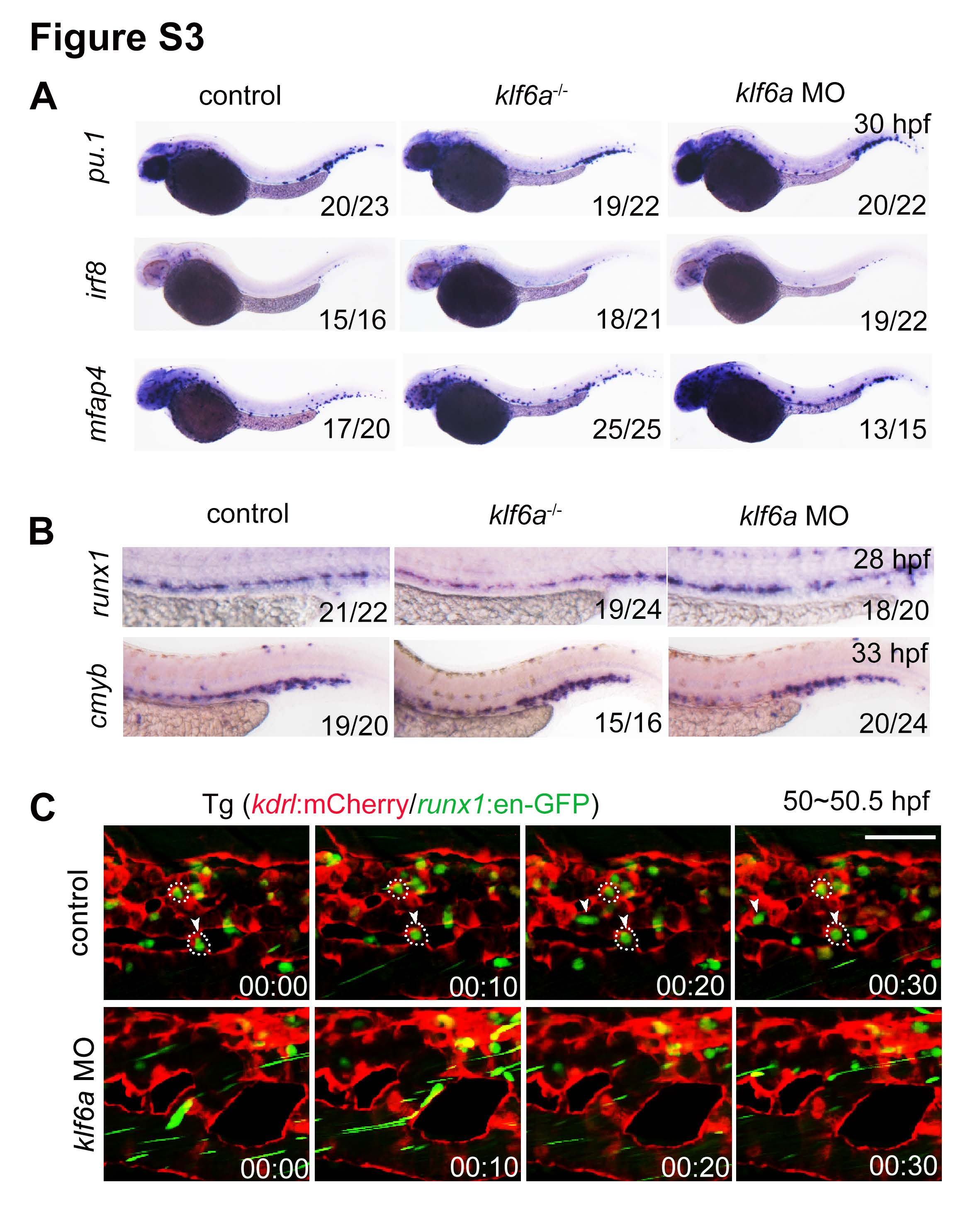
Fig. S3
The blockage of HSPCs seeding into the CHT leads to reduced HSPC expansion. Related to Figure 3.
(A) Expressions of irf8, pu.1 and mfap4 in control and klf6a deficient embryos at 30 hpf. (B) Expression of runx1 and cmyb in control and klf6a deficient embryos at 28 hpf and 33 hpf. (C) Time-lapse confocal imaging of Tg (kdrl:mCherry/runx1:en-GFP) at 50-50.5 hpf in control and klf6a morphants. The arrowheads showing the HSPC lodgement in the CHT vascular niche. The white circles indicate EC pocket. Scale bar, 50 μm.
Acknowledgments
This image is the copyrighted work of the attributed author or publisher, and
ZFIN has permission only to display this image to its users.
Additional permissions should be obtained from the applicable author or publisher of the image.
Reprinted from Developmental Cell, 42(4), Xue, Y., Lv, J., Zhang, C., Wang, L., Ma, D., Liu, F., The Vascular Niche Regulates Hematopoietic Stem and Progenitor Cell Lodgment and Expansion via klf6a-ccl25b, 349-362.e4, Copyright (2017) with permission from Elsevier. Full text @ Dev. Cell