
Image
|
Figure Caption
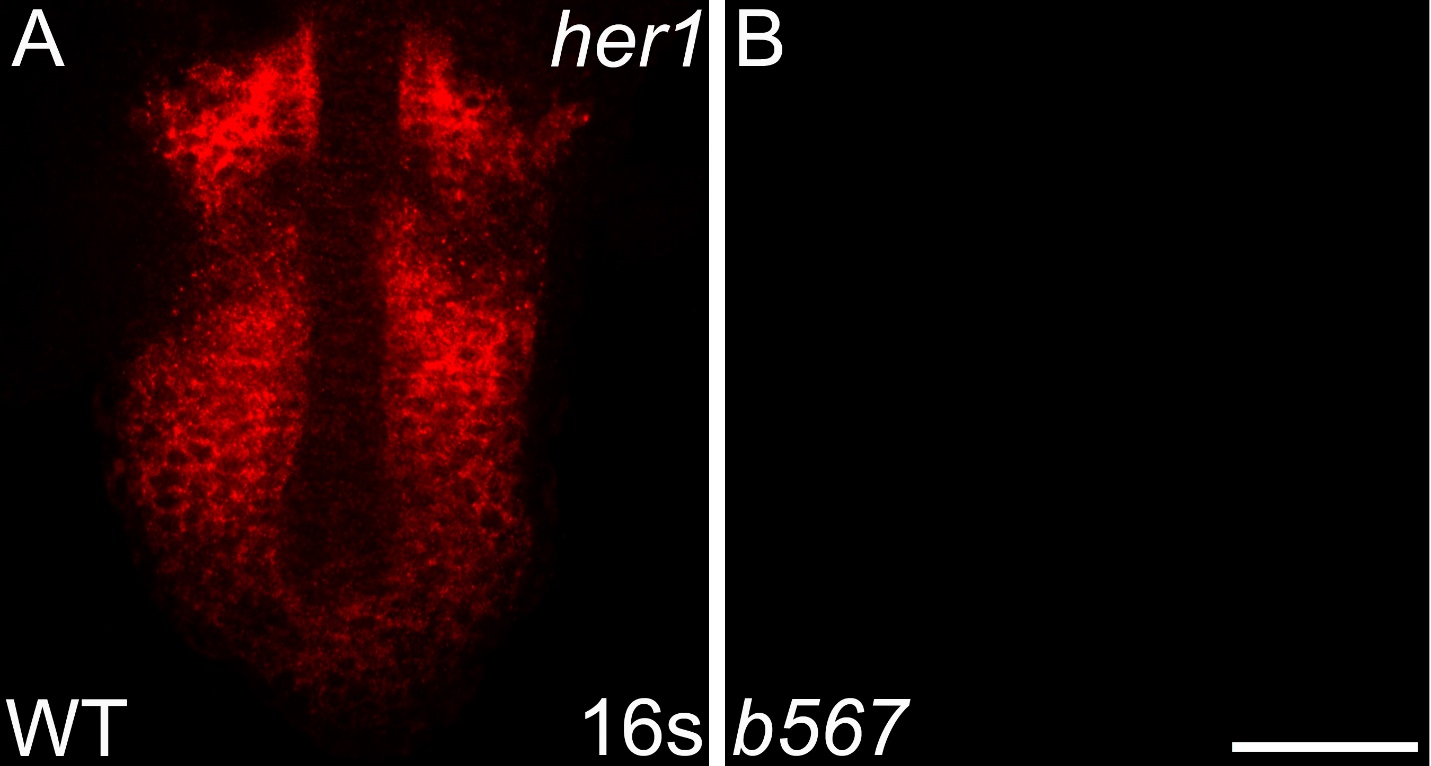
Fig. S5
her1 transcripts are not detected in embryos homozygous for a her1 deficiency. Detection of her1 mRNA by in situ hybridization chain reaction (HCR-ISH) by confocal microscopy reveals striped her1 expression in the presomitic mesoderm (PSM) of wild-type embryos (A), mirroring what is observed by colorimetric in situ. As expected, Df(Chr05:her1,her7,ndrg3a)b567 homozygote embryos, imaged using the same laser intensity and exposure as wild-type embryos in A, lack detectable her1 expression (B). Scale bar = 50 um.
Acknowledgments
This image is the copyrighted work of the attributed author or publisher, and
ZFIN has permission only to display this image to its users.
Additional permissions should be obtained from the applicable author or publisher of the image.
Reprinted from Developmental Biology, 429(1), Gallagher, T.L., Tietz, K.T., Morrow, Z.T., McCammon, J.M., Goldrich, M.L., Derr, N.L., Amacher, S.L., Pnrc2 regulates 3'UTR-mediated decay of segmentation clock-associated transcripts during zebrafish segmentation, 225-239, Copyright (2017) with permission from Elsevier. Full text @ Dev. Biol.