
|
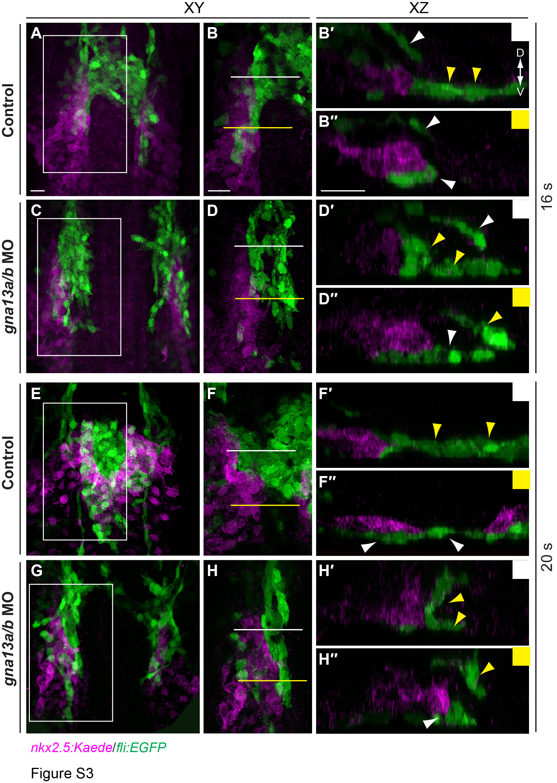
Fig. S3
The relative positions of endocardial and myocardial precursors are disrupted in Gα13-deficient embryos. Confocal images showing endocardial (green) and myocardial (magenta, revealed by Kaede immunostaining) precursors in control and gna13a/b MO-injected embryos at the indicated stages. (A, C, E, G) XY projections of confocal Z-stacks (B, D, F, H) Higher-magnification views of the areas shown in boxes in A, C, E, G. (B'-B'', D'-D'', F'-F'', H'-H'') XZ transverse sections from the areas indicated by white and yellow lines in B, D, F, H. Yellow arrowheads: presumptive endocardial precursors; white arrowheads: endothelial cells. D: dorsal; V: ventral. Scale bars: 20 μm.
Reprinted from Developmental Biology, 414, Xie, H., Ye, D., Sepich, D., Lin, F., S1pr2/Gα13 signaling regulates the migration of endocardial precursors by controlling endoderm convergence, 228-43, Copyright (2016) with permission from Elsevier. Full text @ Dev. Biol.