
|
Fig. S5
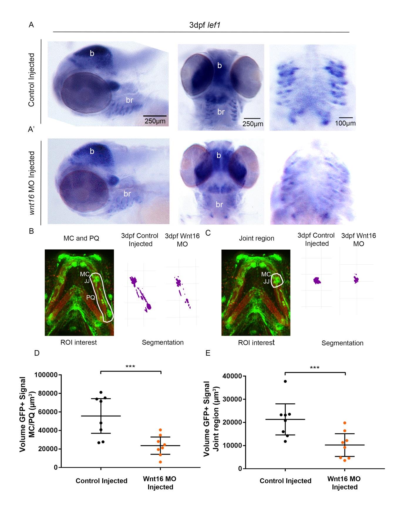
Wnt16 morpholino knockdown affects canonical Wnt activation in the jaw. (A,A’): in situ hybridisation of lef1 mRNA expression in 3dpf control injected (A) and Wnt16 MO injected (A’) zebrafish. (n=9, 19 animals). Left panels: lateral view of head. Middle panels: Ventral view of head. Right panels: Ventral view of branchial arches. B= brain, br=branchial arches. (B): Left panel: volume analysis of Tg(7xTCF.XlaSiam:nlsGFP) GFP-positive (GFP+) signal at the region of interest (ROI) from the Meckel’s Cartilage (MC) jaw joint (JJ) and along the full extent of the palatoquadrate (PQ) (white line). Right panel: Segmentation of GFP+ signal volume from region of interest in 3dpf control injected and Wnt16 morphant zebrafish. (C): Left panel: volume analysis of Tg(7xTCF.XlaSiam:nlsGFP) GFP-positive (GFP+) signal at the ROI from the Meckel’s Cartilage (MC) jaw joint to the interzone (white line). Right panel: Segmentation of GFP+ signal volume from region of interest in 3dpf control injected and Wnt16 morphant zebrafish. (D): Volume (μm3) of GFP+ signal at the MC joint and along the PQ (as measured in (B)) in 3dpf control injected and Wnt16 MO injected zebrafish. (n=8, 8 joints). (E): Volume (μm3) of GFP+ signal at the MC joint (as measured in (C)) in 3dpf control injected and Wnt16 MO injected zebrafish. (n=8, 8 joints). Two-tailed student t- tests were performed for (D,E). ***=p≤0.001. Bars on graph represent mean and 95%CI.