
Image
|
Figure Caption
Fig. 12
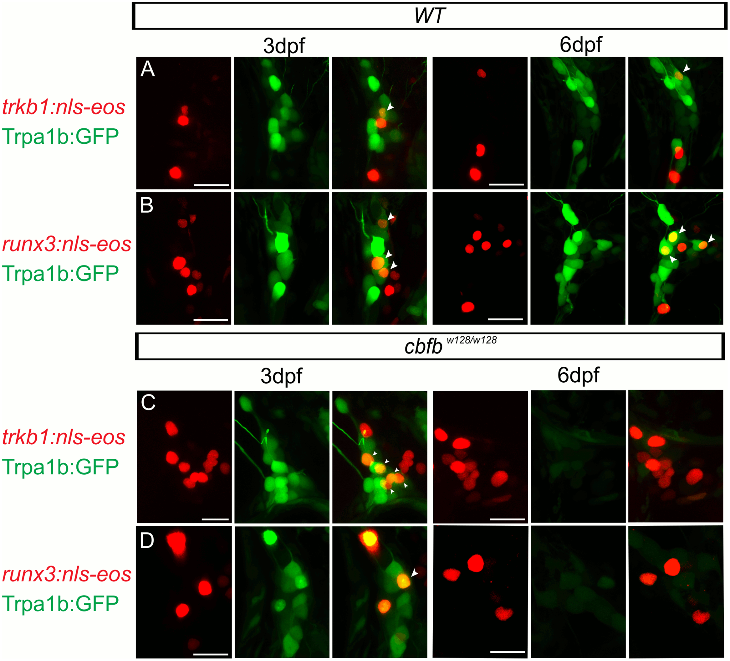
Scatter labeled trkB1 and runx3 neurons persist in cbfbw128/w128; trpa1b:GFP embryos.
(A-D) Maximum intensity projections of transiently expressed nls-Eos in trpA1b:GFP fish at 3dpf and 6dpf. (A) trkb1:nls-Eos in a WT trpa1b:GFP embryo, (B) runx3:nls-Eos in a WT trpa1b:GFP embryo, (C) trkB1:nls-Eos in a in cbfbw128/w128; trpa1b:GFP embryo, (D) runx3:nls-Eos in a in cbfbw128/w128; trpa1b:GFP embryo. Arrowhead indicates double positive nls-Eos/GFP TG neurons. Scale bar: 20μm.
Acknowledgments
This image is the copyrighted work of the attributed author or publisher, and
ZFIN has permission only to display this image to its users.
Additional permissions should be obtained from the applicable author or publisher of the image.
Full text @ PLoS Genet.