
|
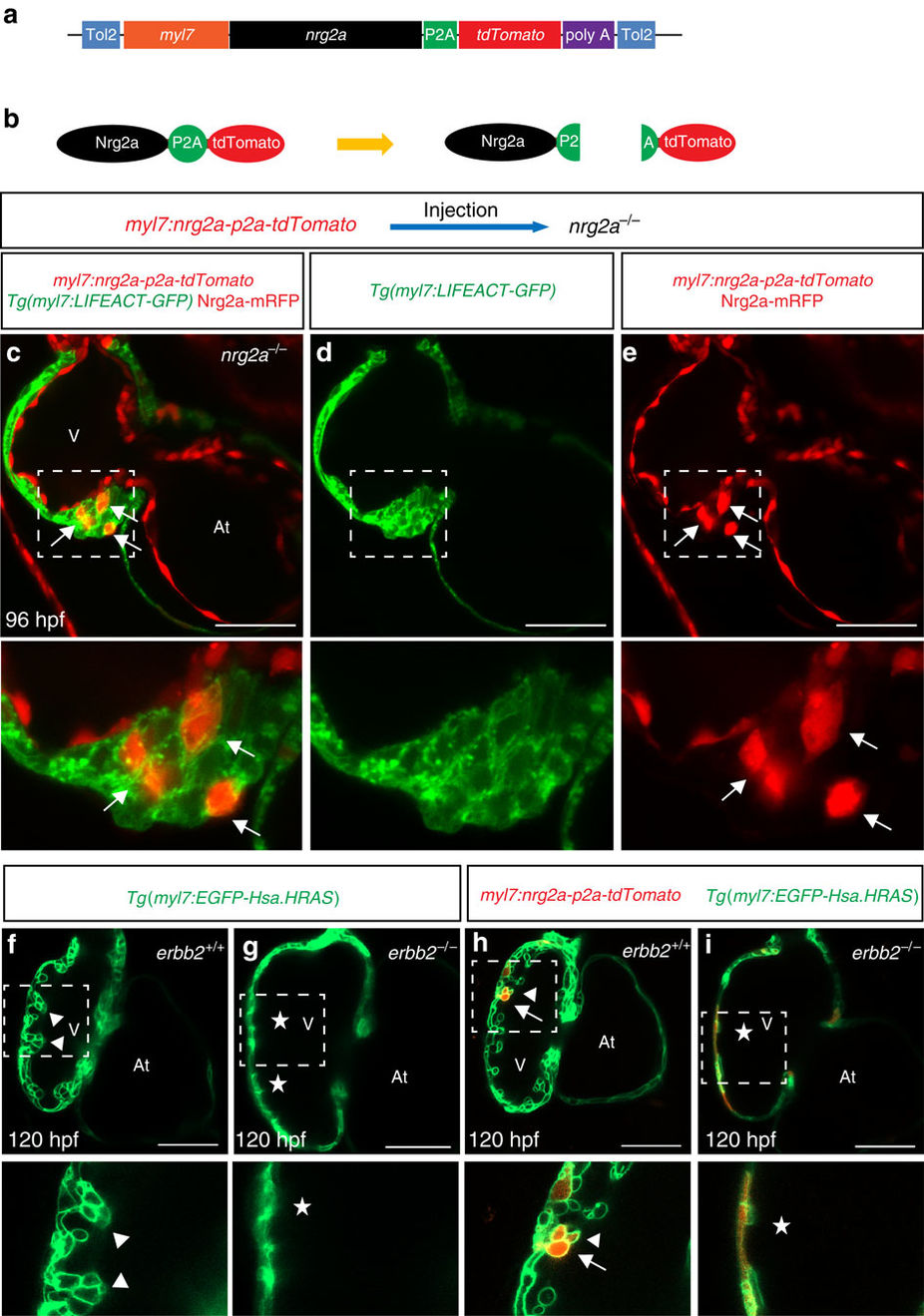
Fig. 4
Myocardial nrg2a overexpression induces cardiomyocyte multilayering in nrg2a mutants.
(a) Cartoon of myocardial specific nrg2a construct. (b) Schematic representation of Nrg2a protein tagged by tdTomato. Due to the presence of the P2A peptide, cleavage occurs right after protein translation to separate the Nrg2a from the tdTomato fluorescent protein. (c–e) 2D confocal images (mid-sagittal sections) of Tg(myl7:LIFEACT-GFP);nrg2a−/− hearts injected with myocardial specific nrg2a construct (myl7:nrg2a-p2a-tdTomato) at the one-cell stage. Mosaic overexpression of nrg2a in nrg2a−/− cardiomyocytes led to the formation of a multilayered myocardial wall which is outlined by a white dashed box and magnified (c–e); arrows point to nrg2a overexpressing cardiomyocytes. (f–i) Confocal images (mid-sagittal sections) of 120 hpf Tg(myl7:EGFP-Hsa.HRAS) hearts from erbb2+/− incrosses injected with the myl7:nrg2a-p2a-tdTomato construct (h–i). Magnified images of dashed boxes are shown below c–i; arrows point to nrg2a overexpressing cardiomyocytes, arrowheads point to trabeculae and asterisks indicate lack of trabeculae. At, atrium; V, ventricle; scale bars, 50 μm.