
|
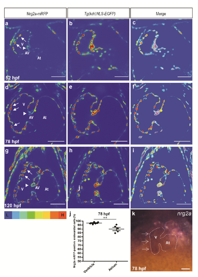
Fig. S2
Cardiac Nrg2a-mRFP expression during embryonic and larval development
(a-i) Mid-sagittal confocal images of Tg(kdrl:NLS-EGFP);nrg2a +/- hearts; heat map; low intensity (blue) to high intensity(red) at 52 (a-c), 78 (d-f) and 120 (g-i) hpf; arrows and arrowheads point to endocardial cells in ventricular outer curvature and superior AV valve leaflet, respectively. (j) Graph showing that on average there is a higher percentage of endocardial cells positive for Nrg2a-mRFP in the ventricle than in the atrium at 78 hpf; dots in this graph represent individual hearts; N=5 hearts; values represent means ± SEM; ** P≤ 0.01 by Student’s t-test. (k) In situ hybridization for nrg2a expression in 78 hpf heart. V: ventricle, AV: atrioventricular canal, At: atrium; scale bars, 50 μm.