
|
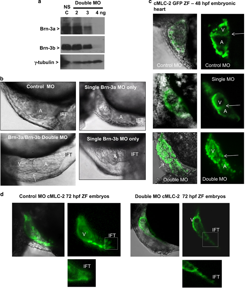
Fig. 5
(a) Western blot analysis showing changes in Brn-3a or Brn-3b proteins extracts prepared from embryos at 72 hpf following injection with different amounts of oligonucleotide MO used to target both Brn-3a and Brn-3b, in fertilised eggs. Gamma (γ) tubulin was used to control for variation in total protein. (b) Photomicrographs showing bright field images of zebrafish heart at 48 hpf following injection of control non-silencing MO only, Brn-3a MO only, Brn-3b MO only or both Brn-3a and Brn-3b (double MO). The failure to loop, which results in linear double morphant heart is indicated by arrow. (c) Representative images showing results of similar studies carried out in CMLC2-GFP, in which the heart is marked with green fluorescent proteins. Left panels show merge of bright field and GFP, and right panels show GFP only in embryonic hearts taken from embryos following injection with control MO, single MO or double MO to target both Brn-3a and Brn-3b. (d) Representative images of hearts from ZF embryos at 72 hpf following injection with control MO or double MO is shown to highlight the significant changes in inflow tract when both Brn-3a and Brn-3b are targeted. A=atria; hpf, hours post fertilisation; IFT, inflow tract; MO, morpholinos; V, ventricle