
|
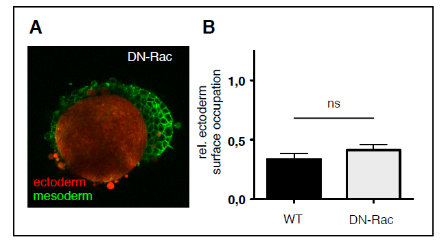
Fig. S4
Envelopment of ectoderm and mesoderm cell aggregates overexpressing DN-rac in vitro. (A) Representative single plane confocal images of tissue aggregates consisting of ectoderm or mesoderm progenitor cells cultured for 5 h in the presence of medium with 300 (A) and 250 mOsm/L osmolarity. Both ectoderm and mesoderm cells express DN-Rac, and Lyn-Venus at the plasma membrane (green). Ectoderm aggregates were additionally labeled with cytoplasmic Dextran-Alexa648 (red). Scale bar, 50 μm. (B) Degree of envelopment was quantified by calculating the relative ectoderm surface occupation taking the heterotypical cell aggregate size into account. Error bars are standard deviations.