
|
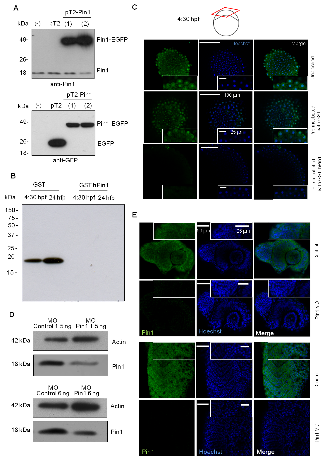
Fig. S2
Pin1 polyclonal antibody specifically recognizes D. rerio Pin1.
(A) Western blot analysis of extracts from HEK-293 cells transfected with pT2-Pin1 or pT2AL500R150G (pT2) probed with Pin1 antibody (upper panel) or GFP antibody (lower panel), (-) untransfected cells, (1) and (2) indicate two independent transfections. (B) Western blot analysis of embryonic extracts from the indicated stages with Pin1 antibody pre-absorbed with recombinant GST or GST-hPin1. (C) Whole-mount immunofluorescence of 4:30 hpf embryos with untreated Pin1 antibody (upper panels), Pin1 antibody pre-absorbed with recombinant GST (middle panels) or pre-absorbed with GST-hPin1 (lower panels). The insets show digital magnifications of selected regions from each image. (D) Zebrafish embryos were microinjected at 1 cell-stage with 1.5 or 6 ng of control or Pin1 specific morpholinos (MO), and upon 24 hours, western blot was performed on protein extracts using anti Pin1 and anti Actin as loading control (E) Whole-mount immunofluorescence of 6 ng Pin1 MO or control MO microinjected embryos at 24 hpf using Pin1 antibody, showing part of the head (upper panels), or trunk (lower panels). The insets show digital magnifications of selected regions from each image.