
|
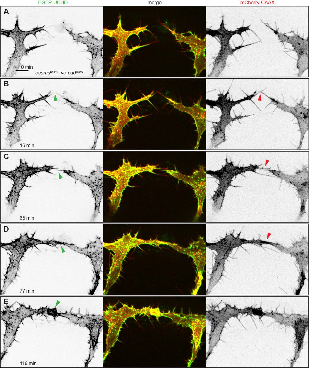
Fig. S4
esama-/-; ve-cad+/- embryos display an intermediate filopodial phenotype. (A-F) Still images from Movie S9 of an esamaubs19; ve-cad+/ubs8 embryo. Tg(fli1ep:gal4ff)ubs3, Tg(UAS:EGFP-UCHD)ubs18, Tg(kdrl:mCherry-CAAX)s916 embryo at around 32hpf, anterior to the left. Single channels are shown in inversed contrast (green is EGFP-UCHD and red is mCherry-CAAX on left and right, respectively) and the merge is shown in the middle. (A) Two tip cells are extending filopodia towards each other. (B) Two filopodia touch (red arrowhead). (C) A filopodial contact is established (red arrowhead) and actin cytoskeleton (green arrowhead) is being recruited to the cell-cell contact site. (D) The cell-cell bridge (red arrowhead) is quickly stabilized with actin cytoskeleton (green arrowhead). (E) Anastomosis led to the formation of a junctional ring between the two tip cells (green arrowhead). Scale bar, 10μm.