
Image
|
Figure Caption
Fig. S1
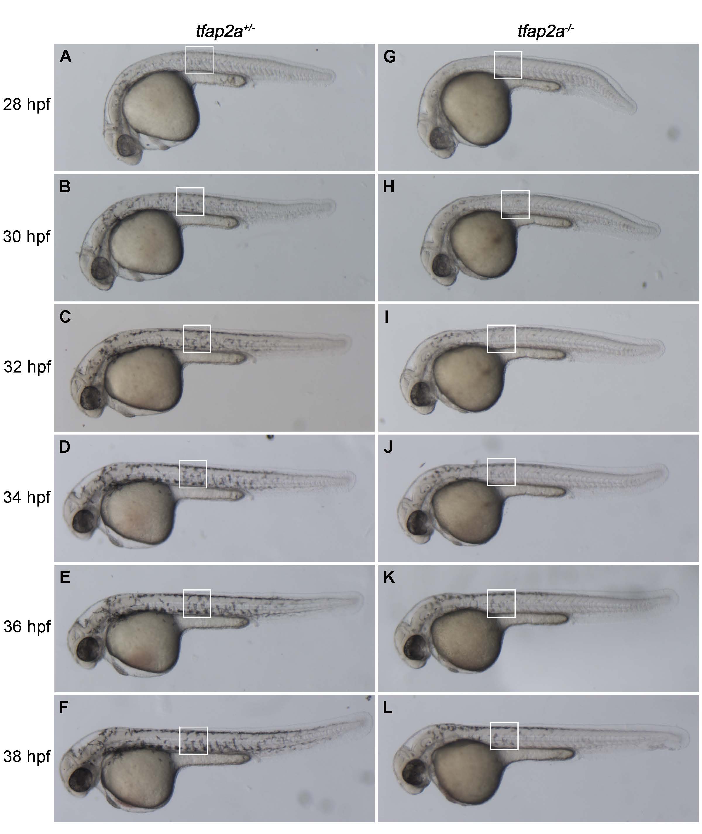
Time course of delayed melanocyte differentiation in tfap2a-/- zebrafish.
(A-L) Lateral views of live tfap2a+/- (left column) and tfap2a-/- (right column) zebrafish embryos at the indicated age from 28-38 hpf. (C, I) Melanocytes in a tfap2a+/- zebrafish (C) appear darkly pigmented by 32 hpf, whereas melanocytes at the same location in a tfap2a-/- mutant (I) at this stage remain pale and punctate. (F, L) At 38 hpf, there is still a detectable difference in the level of pigmentation in tfap2a+/- (F) and tfap2a-/- (L) mutant animals.
Acknowledgments
This image is the copyrighted work of the attributed author or publisher, and
ZFIN has permission only to display this image to its users.
Additional permissions should be obtained from the applicable author or publisher of the image.
Full text @ PLoS Genet.