
|
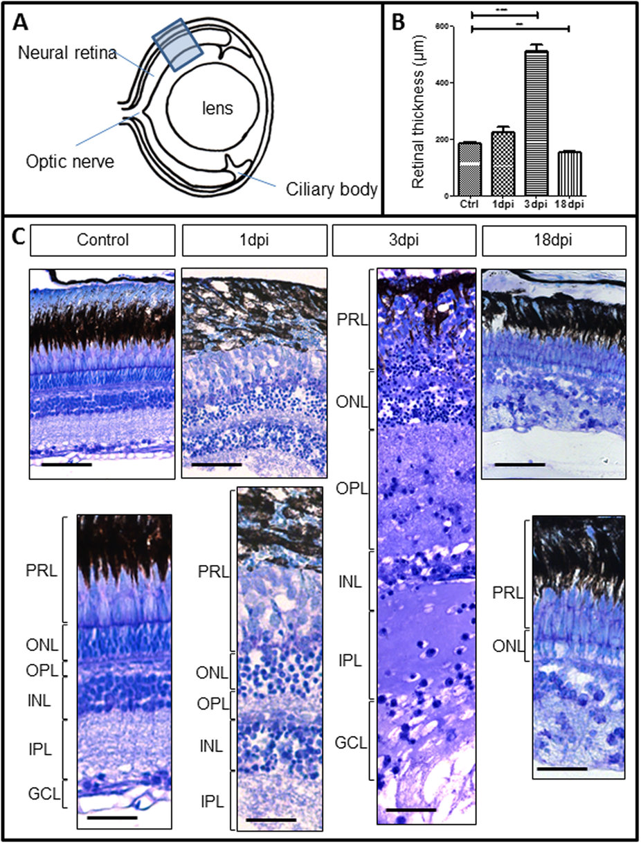
Fig. 1
Comparison of zebrafish retinal structure between normal and Oubain damaged retina at 1, 3 and 18 days post injection (dpi).
(A) Site of retinal sampling from which images in (C) were taken for consistency. (B) Histogram shows the differences in the mean neural layer thickness between control, 1 dpi, 3 dpi and 18 dpi (**P = 0.0044; ***P < 0.0001) n = 3. (C) Retinal sections were stained with toluidine blue. While the retina structure at 1 dpi appears less dense in comparison to control, the retina at 3 dpi almost doubled in size and cellularity was clearly decreased. By 18 dpi the photoreceptor layer had regained structure but the inner retinal layers could not be determined. PRL = photoreceptor layer; ONL = outer nuclear layer; OPL = outer plexiform layer; INL = inner nuclear layer; IPL = inner plexiform layer; GCL = ganglion cell layer. Scale = 50 μm.