
|
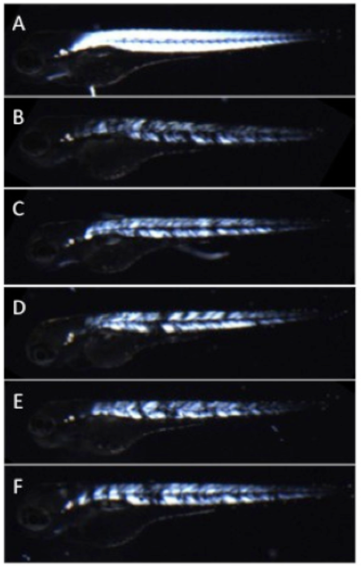
Fig. 2
Muscle fiber detachment of caf mutants can be observed with birefringence.
Under plane polarized light, muscle from WT siblings (A) appears uniformly bright and white, consistent with normal muscle organization. In contrast, Caf mutants (B-F_ can be identified as having stochastic patterns of muscle degeneration and detachment with birefringence as early as 2 dpf. This is seen in the muscle compartment as dim white areas (thinned or atrophied fibers) and black spots (presumed areas of muscle fiber detachment). Of note, genotype for all depicted animals was confirmed by Sanger sequencing. Bars represent mean ± SEM.