
|
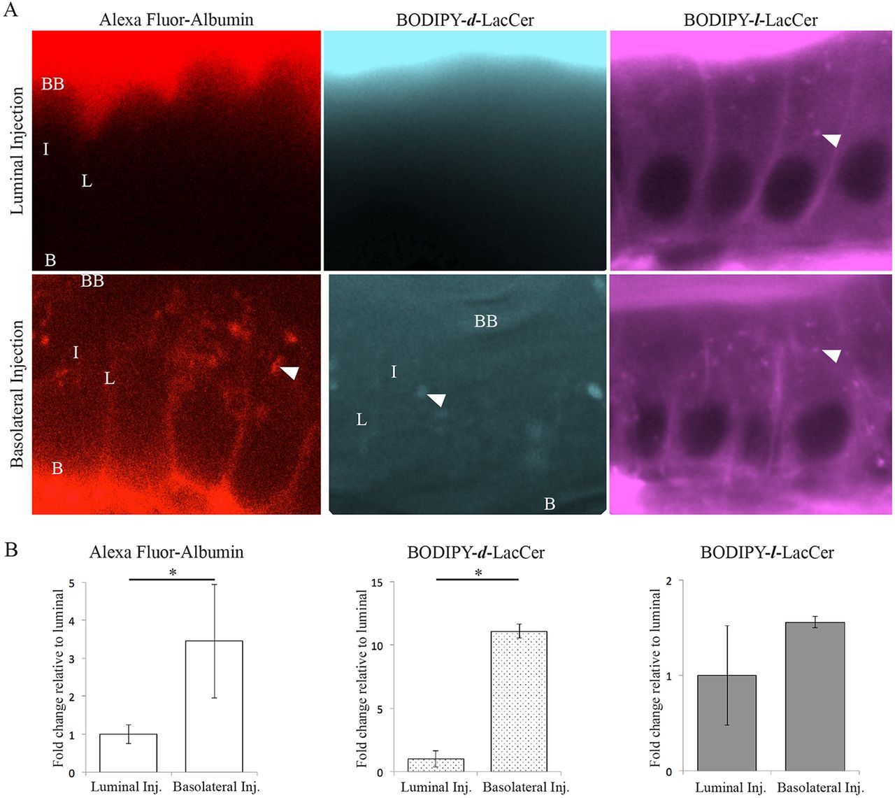
Fig. 2
Fluorescently labeled endocytic cargos enable imaging of caveolar endocytosis in the intact zebrafish intestine. (A) Representative images show that the caveolar-specific cargos Alexa Fluor–albumin and BODIPY–d-LacCer are internalized from the basolateral PM of enterocytes, but not the intestinal lumen. In contrast, the cargo transported specifically by clathrin-coated vesicles, BODIPY–l-LacCer, is transported into enterocytes from both the basolateral and luminal PMs. BB, brush border; L, lateral membrane; B, basolateral membrane; I, intracellular; N, nucleus; arrowhead, intracellular puncta. (B) The mean fluorescence intensity of Alexa Fluor–albumin and BODIPY–d-LacCer on the lateral PM of enterocytes is significantly greater following basolateral injection compared to luminal injection. In contrast, the mean fluorescence intensity of BODIPY–l-LacCer on the lateral PM of enterocytes is the same following basolateral and luminal injection. Data is presented relative to lateral PM mean fluorescence intensity following luminal injection. Mean±s.e.m, n=3: nine fish per replicate, three areas of each region per fish; Student's t-test; *P<0.05.