
|
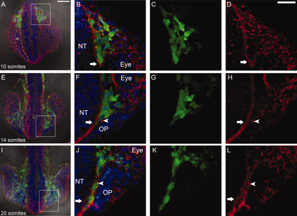
Fig. 8
Anterior migration of neural crest is accompanied by changes in the basal lamina. A,E,I: Whole-mount heads in dorsal view at 10 somite stage (ss) (A), 14 ss (E), and 20 ss (I). Boxed area show region of interest shown at higher magnification. A: Region of cells that will give rise to olfactory placode (OP) indicated by dashed outline (op). B–D: The leading edge of Sox10:GFP positive CNC (green) moves as a group surrounded by laminin (red, arrow). F–H: The cranial neural crest (CNC) (green) passes anterior separating the lamina of the neural tube (NT, red, arrow) and OP (red, arrowhead). J-L: CNC (green) is now flanked by laminin on both the side of the NT (red, arrow) and OP (red, arrowhead). Scale bars = 50 µm in A,E,I; 25 µm in B–D,F–H,J–L.